Disclosure
This book was created to help patients and families better understand neurological conditions in clear and simple language. Artificial intelligence tools were used to organize information and improve readability, always under the guidance and review of the author. All medical explanations, examples, and recommendations have been carefully verified to ensure accuracy and relevance for patient education.
Introduction
Neurology, the study of the brain and nervous system, can often seem overwhelming for patients and families. Medical terms may sound complex, and understanding what is happening in the body can be confusing. This book was created to make neurological conditions easier to understand, using clear and simple explanations that bridge medical knowledge with patient education.
This book is designed as a companion to the A to Z Neurology for Physicians textbook. It follows the same structure but focuses entirely on individual neurological diseases, explained in everyday language. Physicians can use this book as an educational tool to help patients and their families understand specific conditions discussed during clinical visits. It also serves as a helpful guide for anyone who wants to learn more about neurological disorders in a concise and approachable way.
While this book simplifies many medical ideas for easier understanding, it does not replace professional medical advice or serve as a comprehensive reference. Readers are encouraged to consult qualified healthcare professionals and verified medical resources for further information and personalized guidance.
By improving understanding of neurological conditions, this book aims to empower patients and caregivers to take a more active role in their healthcare journey and foster better communication between doctors and patients.
Acute Disseminated Encephalomyelitis (ADEM)
What is it?
ADEM is a short-term inflammation of the brain and spinal cord that usually appears after an infection.
It happens when the body’s defense system mistakenly attacks the brain’s protective coating.
The condition causes temporary problems with thinking, balance, and muscle control.
ADEM most often affects children and young adults.
It usually occurs only once, and most people make a full recovery.
What causes it?
ADEM often develops one to three weeks after a viral or bacterial infection.
The immune system becomes confused and attacks healthy nerve tissue.
Rarely, it may follow a vaccination, though this is uncommon.
The exact cause isn’t fully understood, but it involves an overactive immune response.
It’s not contagious—you can’t spread it to others.
How is it diagnosed?
Doctors consider recent illness, symptoms, and examination findings.
An MRI scan shows small spots of inflammation in different parts of the brain and spinal cord.
A spinal tap can check for signs of infection or inflammation.
Blood tests help rule out other causes such as multiple sclerosis.
The combination of test results and symptoms confirms the diagnosis.
What is the treatment?
High-dose steroids are given by vein to reduce swelling and calm the immune system.
If steroids don’t work, treatments like plasma exchange or IV immunoglobulin can be used.
Supportive care includes rest, fluids, and physical or occupational therapy.
Seizures, headaches, or fever are treated as needed.
Most people start to feel better within days of treatment.
What is the prognosis?
Most patients recover fully within weeks to months.
A few may have mild lasting weakness or coordination issues.
Recurrence is rare.
Regular follow-up helps ensure the brain heals completely.
Long-term outlook is excellent in most cases.
Acute Inflammatory Demyelinating Polyradiculoneuropathy (AIDP)
What is it?
AIDP is a nerve disorder that causes sudden weakness starting in the legs and moving upward.
It’s the most common form of Guillain–Barré syndrome.
The immune system mistakenly attacks the nerves’ protective covering.
This slows down or blocks the messages between the brain and the muscles.
With proper treatment, most people recover well.
What causes it?
It often begins one to three weeks after an infection such as a cold or diarrhea.
The immune system becomes overactive and attacks nerve tissue instead of germs.
Certain bacteria and viruses are more likely to trigger it.
It’s not inherited or contagious.
The exact reason why it happens to some people is not fully known.
How is it diagnosed?
Doctors look for symmetrical weakness that begins in the legs and loss of reflexes.
A spinal tap shows high protein levels but very few cells in the fluid.
Nerve tests show slower electrical signals due to nerve damage.
MRI scans may show swelling of the nerve roots in the spine.
Other similar conditions are ruled out through testing.
What is the treatment?
Hospital care is needed to watch breathing, heart rhythm, and blood pressure.
IV immunoglobulin (IVIG) or plasma exchange helps calm the immune system.
Steroids are not useful and are not recommended.
Physical therapy helps prevent stiffness and improves muscle recovery.
Supportive care, including nutrition and emotional support, aids recovery.
What is the prognosis?
Most people recover strength within a few months.
Some may have mild lingering weakness or fatigue.
Early treatment leads to a faster and more complete recovery.
Severe cases may take longer but usually improve with rehabilitation.
The condition rarely returns once it has resolved.
Alcohol and Neurology
What is it?
Long-term heavy drinking can harm the brain and nerves, leading to balance, memory, and movement problems.
Alcohol affects how nerve cells talk to each other and can shrink certain brain areas.
Common effects include poor coordination, confusion, numbness in the feet or hands, and trouble walking.
Some people develop specific conditions such as Wernicke-Korsakoff syndrome or cerebellar degeneration.
The brain changes are often partly reversible if alcohol use stops early.
What causes it?
Alcohol itself is toxic to nerve and brain cells when used over many years.
Drinking heavily often leads to poor diet and vitamin B1 (thiamine) deficiency, which damages the brain.
Low thiamine levels cause Wernicke’s encephalopathy and memory loss (Korsakoff’s syndrome).
The cerebellum, which controls balance, is especially sensitive to alcohol damage.
Other toxins and liver problems from alcohol can worsen brain function.
How is it diagnosed?
Doctors look for symptoms such as unsteady walking, slurred speech, or numbness in the limbs.
They ask about drinking habits and check for signs of poor nutrition.
Blood tests can show low vitamin levels and liver changes from long-term drinking.
MRI scans may show shrinkage in the cerebellum or other parts of the brain.
Nerve tests can reveal damage to the peripheral nerves in the hands and feet.
What is the treatment?
The most important step is stopping alcohol use completely.
High-dose vitamin B1 (thiamine) is given right away, especially before receiving any sugar or glucose.
Eating a balanced diet with vitamins helps the brain and nerves heal.
Physical and occupational therapy can improve coordination and movement.
Counseling and support groups help maintain sobriety and emotional recovery.
What is the prognosis?
If alcohol use stops early, many symptoms can improve or even disappear.
Memory loss and nerve damage may be partly reversible, though some effects can last.
Continued drinking leads to worsening brain and nerve injury.
Early vitamin replacement can prevent Wernicke-Korsakoff syndrome from becoming permanent.
Long-term recovery depends on full abstinence, good nutrition, and consistent follow-up.
Alzheimer’s Disease
What is it?
Alzheimer’s disease is a brain condition that slowly causes problems with memory and thinking.
It is the most common cause of dementia in older adults.
Over time, people may forget names, places, and daily routines.
The disease affects how brain cells communicate and eventually leads to their loss.
It develops gradually, starting with forgetfulness and progressing to more severe confusion.
What causes it?
The disease happens when harmful proteins build up in the brain and damage nerve cells.
These proteins—called amyloid and tau—disrupt brain cell connections.
Age, family history, and certain genes increase the risk.
Poor heart health, diabetes, and head injuries can also raise the risk.
The exact cause isn’t fully known, but lifestyle and genetics both play roles.
How is it diagnosed?
Doctors start by asking about memory, mood, and daily function.
Simple tests check thinking, attention, and problem-solving abilities.
MRI or CT scans can show shrinkage in memory-related brain areas.
Blood and spinal fluid tests help rule out other causes of memory loss.
Early diagnosis allows better planning and support for the patient and family.
What is the treatment?
Medicines like donepezil or memantine can help with memory and behavior.
Staying mentally and socially active slows down memory decline.
A healthy diet, regular exercise, and good sleep support brain health.
Counseling and support groups help families manage daily challenges.
New treatments are being developed to target the brain changes directly.
What is the prognosis?
Alzheimer’s disease usually worsens slowly over several years.
Early stages involve forgetfulness, while later stages affect all daily activities.
With care and treatment, many people can live meaningful lives for years.
The focus of treatment is comfort, safety, and quality of life.
Support from caregivers and medical teams makes a major difference in long-term well-being.
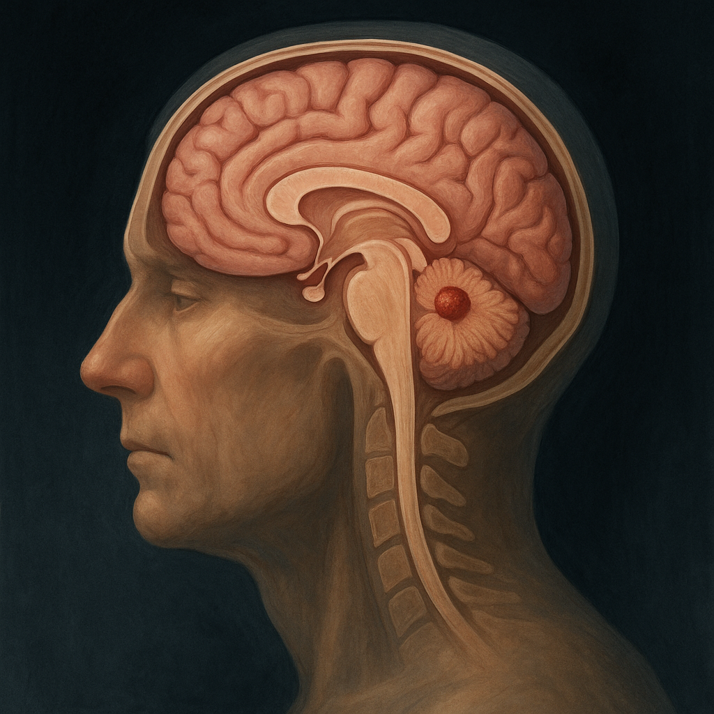
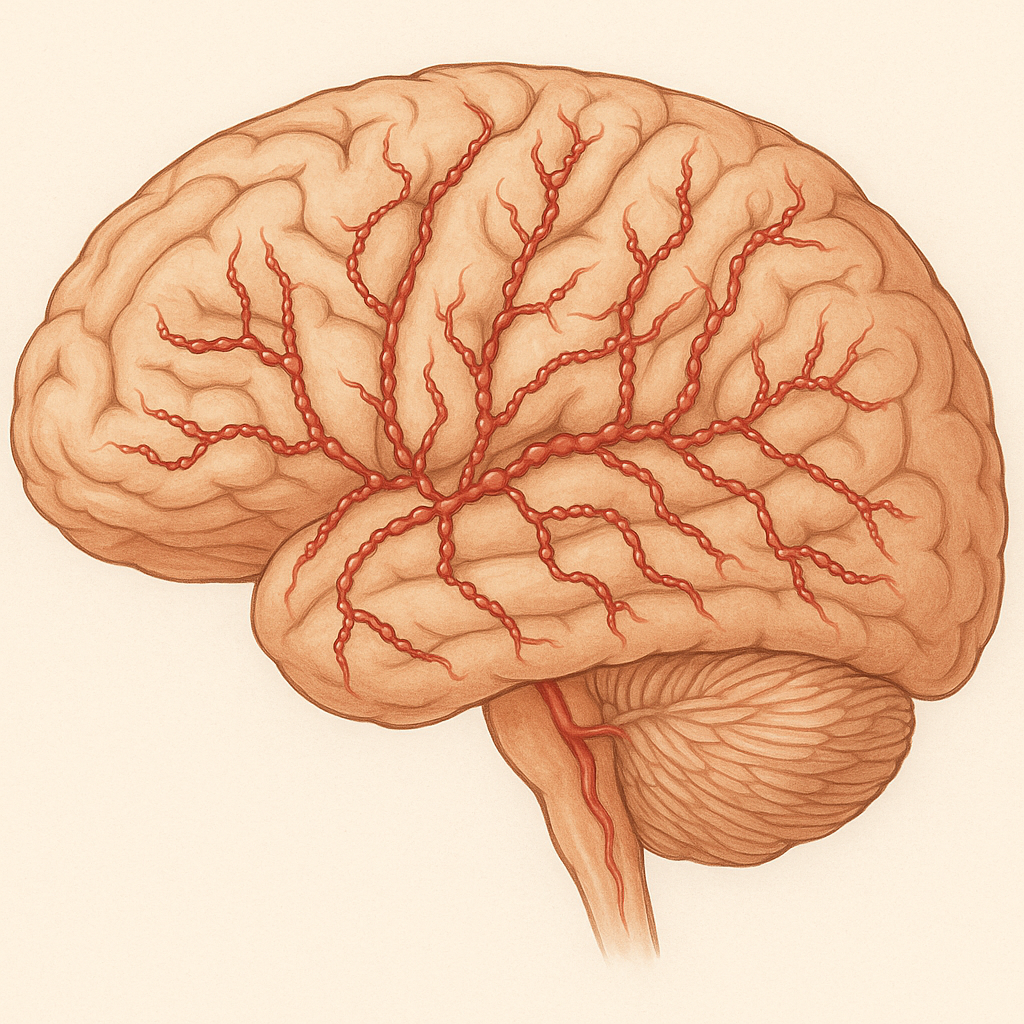
Amyloid Angiopathy
What is it?
Amyloid angiopathy (CAA) is a condition in which abnormal protein called amyloid-\(\beta\) builds up in the walls of small blood vessels in the brain.
This weakens the vessels, making them more likely to bleed and causing lobar intracerebral hemorrhages.
It mainly affects older adults and is a common cause of spontaneous (non-hypertensive) brain bleeding.
The same protein is also involved in Alzheimer’s disease, linking the two conditions.
CAA can also cause small “silent” bleeds, cognitive changes, or transient neurological symptoms.
What causes it?
The disease is caused by gradual deposition of amyloid-\(\beta\) (especially A\(\beta\)40) in the walls of cortical and leptomeningeal arteries.
These deposits damage and weaken the vessel wall, leading to bleeding.
The underlying process relates to problems with amyloid clearance from the brain.
Genetic factors such as APOE \(\varepsilon\)2 and \(\varepsilon\)4 increase risk.
It is not caused by high blood pressure, trauma, or infection.
How is it diagnosed?
MRI scans show typical findings — multiple small “microbleeds” in the cortex and cortical superficial siderosis (thin lines of old blood on the surface).
CT scans help detect acute bleeds, usually in the lobar (outer) brain regions.
The diagnosis is made using the Boston Criteria, based on clinical and MRI findings.
A brain biopsy is rarely needed, only if other causes like vasculitis are suspected.
Other conditions such as hypertensive bleeds or vascular malformations must be ruled out.
What are the symptoms?
Sudden weakness, speech disturbance, or confusion due to lobar hemorrhage.
Recurrent episodes of lobar bleeding over time.
Progressive memory loss and cognitive decline in some patients.
Transient focal neurological episodes (TFNEs) — short, stroke-like spells that fully recover.
Less commonly, seizures or headaches during acute bleeds.
What is the treatment?
There is no cure, but treatment aims to prevent further bleeding and manage symptoms.
Strict blood pressure control is essential to reduce recurrence risk.
Avoid blood thinners and antiplatelet medications if possible.
Seizures are treated with appropriate antiepileptic drugs.
Physical and cognitive rehabilitation can help recovery after bleeds.
Experimental therapies targeting amyloid clearance are under study.
What is the prognosis?
The outlook depends on the severity and recurrence of bleeding.
Many patients recover from a single bleed, but recurrent episodes can lead to lasting disability or dementia.
Avoiding risk factors and close medical follow-up help reduce complications.
Long-term management focuses on maintaining independence and preventing further hemorrhage.
Supportive care and monitoring for cognitive symptoms improve quality of life.
Amyloid Neuropathy
What is it?
Amyloid neuropathy is a nerve problem caused by abnormal protein deposits called amyloid.
These proteins build up in and around nerves, affecting their ability to send signals.
It often causes tingling, burning, or numbness in the feet and hands.
Some people also have dizziness, stomach problems, or trouble controlling blood pressure.
The condition can be inherited or develop later in life as part of another illness.
What causes it?
It occurs when certain proteins fold the wrong way and form sticky deposits that damage nerves.
These proteins can come from genetic changes (inherited) or from disorders of blood cells.
In some people, long-term inflammatory diseases can also lead to amyloid buildup.
The deposits block blood supply and nutrition to nerve fibers.
Over time, this causes nerve fibers to die and symptoms to progress.
How is it diagnosed?
Doctors suspect the diagnosis when there is numbness, pain, and dizziness that worsen slowly.
A nerve test can show how well the nerves are working and reveal damage.
A small biopsy of fat or nerve tissue confirms amyloid by special staining under the microscope.
Blood and genetic tests help find the type of amyloid and guide treatment.
Scans or heart and kidney tests may check if other organs are also affected.
What is the treatment?
Treatment focuses on stopping more amyloid from forming and relieving symptoms.
Inherited forms may be treated with new medicines that block or silence the abnormal protein.
For acquired cases, chemotherapy or stem cell therapy may remove the source of amyloid.
Pain medicines, blood pressure support, and diet changes help manage daily symptoms.
Early treatment and healthy lifestyle changes can slow or even improve nerve damage.
What is the prognosis?
With early diagnosis and proper treatment, many people can control the disease.
Some nerve damage may improve once amyloid production is stopped.
Delayed treatment can lead to permanent weakness and loss of sensation.
The outcome depends on the type of amyloid and how much other organs are involved.
Regular follow-up helps track progress and adjust therapy as needed.
Amyotrophic Lateral Sclerosis (ALS)
What is it?
ALS is a disease that affects the nerve cells that control muscles.
These nerves gradually stop working, causing weakness and muscle wasting.
It can start in the hands, feet, or speech muscles and slowly spread to other areas.
The condition does not affect thinking or senses in most people.
Over time, the muscles that help with breathing and swallowing also weaken.
What causes it?
Most cases occur without a clear cause, but about one in ten is inherited.
Changes in certain genes can make nerve cells more sensitive to damage.
Chemicals that overstimulate or stress nerve cells may play a role.
Inflammation and poor energy supply to the nerves also contribute.
The condition is not caused by infections or lifestyle choices.
How is it diagnosed?
Doctors diagnose ALS based on the pattern of weakness and muscle changes.
Nerve and muscle tests (EMG and NCS) show how well the nerves send signals.
MRI scans help rule out other conditions like spinal cord problems.
Blood and genetic tests may be used to confirm or exclude inherited forms.
A neurologist experienced in motor neuron diseases usually confirms the diagnosis.
What is the treatment?
There is no cure, but medicines like riluzole and edaravone can slow disease progression.
Breathing and swallowing support are key parts of care as symptoms advance.
Physical, speech, and occupational therapy help maintain mobility and communication.
Nutrition and emotional support improve comfort and quality of life.
New research is exploring treatments that target the disease at a genetic level.
What is the prognosis?
ALS progresses at different rates in different people.
Many people live for several years after diagnosis, and some live much longer.
Early multidisciplinary care improves both comfort and survival.
Breathing support can greatly extend life expectancy.
Ongoing research gives hope for better treatments and possible future cures.
Anoxic Brain Injury
What is it?
Anoxic brain injury happens when the brain does not get enough oxygen for several minutes.
It often occurs after a cardiac arrest, drowning, or choking.
Without oxygen, brain cells begin to die quickly, especially in areas that control memory and movement.
Some people recover partially, while others may remain in a coma or have lasting disabilities.
The severity depends on how long the brain went without oxygen and how quickly help arrived.
What causes it?
The main cause is a complete stop or severe drop in oxygen supply to the brain.
Common triggers include cardiac arrest, suffocation, severe asthma, or drug overdose.
Stroke, carbon monoxide poisoning, or electrical shock can also cause it.
When blood flow stops, brain cells lose energy and begin to break down.
Even after oxygen returns, inflammation and toxins can cause more damage.
How is it diagnosed?
Doctors suspect it when a person does not wake up after oxygen loss.
Brain scans such as MRI or CT show which areas are damaged.
EEG tests measure brain activity and can help predict recovery chances.
Blood tests may show signs of stress or injury to brain cells.
Doctors use all this information together to understand the extent of the injury.
What is the treatment?
The first step is restoring oxygen and blood flow as soon as possible.
Cooling the body (targeted temperature therapy) can help reduce further brain damage.
Doctors control seizures, maintain normal blood pressure, and prevent infections.
Once stable, rehabilitation helps with movement, speech, and memory recovery.
Family support and therapy are key for long-term improvement and quality of life.
What is the prognosis?
Recovery depends on how long the brain went without oxygen and the amount of damage.
Some people may wake up and recover slowly over weeks or months.
Others may have lasting problems with memory, speech, or movement.
Severe cases may lead to coma, vegetative state, or death.
Early resuscitation and care greatly improve the chances of recovery.
Anterior Interosseous Nerve Syndrome (AINS)
What is it?
AINS is a nerve problem that causes weakness in certain hand and forearm muscles.
It affects a small branch of the median nerve that controls thumb and index finger movement.
People with AINS may have trouble making the “OK” sign with their fingers.
It usually causes weakness but no numbness or tingling.
The condition often improves on its own but may take several months.
What causes it?
It can happen when the nerve is squeezed by tight muscles or tendons in the forearm.
Sometimes it follows a viral infection or inflammation of the nerve (neuritis).
Overuse or repetitive arm movement may contribute in some cases.
Rarely, trauma or surgery near the elbow can damage the nerve.
In many people, the exact cause is never found.
How is it diagnosed?
Doctors look for weakness in the thumb and index finger without loss of feeling.
The “pinch test” helps show the classic triangular “OK” sign weakness.
Nerve and muscle tests (EMG) confirm which muscles are affected.
MRI or ultrasound may be done to check for nerve compression or swelling.
The diagnosis is mostly clinical, based on symptoms and physical exam.
What is the treatment?
Most cases improve naturally over time without surgery.
Rest, gentle stretching, and physical therapy help recovery.
Pain and inflammation can be managed with medications if needed.
If the nerve does not recover after 6–12 months, surgery may relieve pressure.
Recovery is usually good, and most people regain near-normal hand strength.
What is the prognosis?
Many patients recover fully within 3 to 6 months.
Some may have mild, lasting weakness in fine finger movements.
Early physical therapy helps prevent stiffness and speed up healing.
Inflammation-related cases respond faster than those caused by compression.
Overall, the long-term outlook is excellent for most people.
Arachnoiditis
What is it?
Arachnoiditis is a long-term inflammation of one of the coverings around the spinal cord called the arachnoid membrane.
It causes thickening and scarring that can trap and irritate the spinal nerves.
People with this condition often have severe, burning pain in the lower back and legs.
It can also affect bladder and bowel control in some people.
Arachnoiditis is chronic, meaning symptoms may last for years and need ongoing care.
What causes it?
The most common cause is irritation from spinal surgery, injections, or infections.
Past spinal procedures, especially epidural or intrathecal injections, can lead to inflammation.
Rare causes include trauma, bleeding around the spinal cord, or chemical irritation.
Sometimes infections like tuberculosis or syphilis can trigger it.
In many cases, a combination of surgical and inflammatory factors is involved.
How is it diagnosed?
Doctors suspect it when back and leg pain persist after surgery or spinal injections.
MRI scans show scarring, thickened tissues, and clumping of nerve roots.
Nerve tests may show changes in multiple nerve roots, though they are not specific.
A detailed history of past spine procedures helps confirm the diagnosis.
Other conditions like spinal stenosis or nerve injury must be ruled out.
What is the treatment?
There is no cure, but treatments can ease pain and improve function.
Medications like gabapentin, pregabalin, or duloxetine help reduce nerve pain.
Physical therapy and gentle stretching improve mobility and strength.
In some cases, spinal cord stimulators or pain management programs are helpful.
Surgery is rarely done, as it may worsen scarring, but may help selected patients.
What is the prognosis?
The condition is long-term but varies from person to person.
Many patients live with chronic pain that can be managed with treatment.
Early diagnosis and a team approach improve comfort and function.
Emotional support and counseling can help with coping and quality of life.
Regular follow-up helps adjust therapy as symptoms change over time.
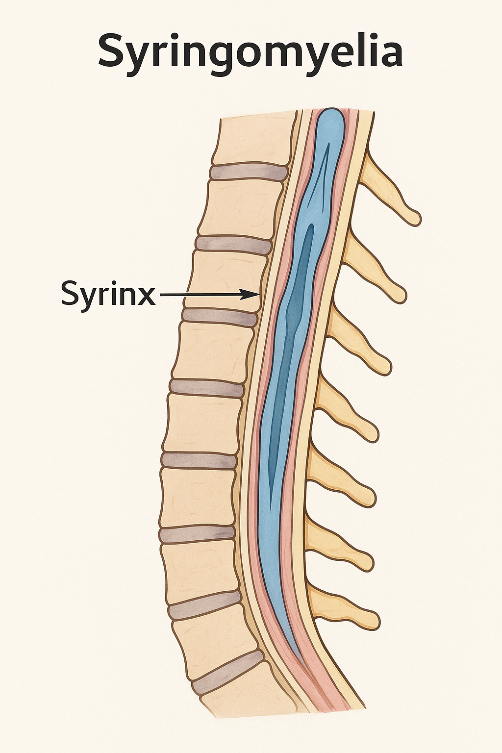
Arsenic Toxicity
What is it?
Arsenic toxicity occurs when the body is exposed to excessive amounts of arsenic, a naturally occurring element found in groundwater, industrial emissions, and contaminated food sources.
Chronic exposure most commonly affects the skin, peripheral nerves, and internal organs.
It can lead to neurological, dermatological, and systemic manifestations.
Arsenic exists in both organic and inorganic forms, with the inorganic type being more toxic.
Long-term exposure is a major global health concern, especially in areas with contaminated well water.
What causes it?
Chronic exposure to arsenic-contaminated groundwater is the most common cause worldwide.
Occupational exposure occurs in industries such as mining, smelting, and pesticide manufacturing.
Arsenic binds to sulfhydryl groups and disrupts cellular respiration, impairing mitochondrial energy metabolism.
It generates reactive oxygen species, leading to oxidative stress, DNA damage, and apoptosis.
Genetic and nutritional factors can modify susceptibility to arsenic toxicity.
How is it diagnosed?
Diagnosis is based on clinical presentation, exposure history, and laboratory testing.
Urine arsenic measurement is the most reliable indicator of recent exposure.
Hair and nail arsenic levels reflect long-term exposure.
Nerve conduction studies show length-dependent axonal neuropathy, especially involving sensory fibers.
Water and environmental testing help identify the source of contamination.
What are the symptoms?
Tingling, numbness, and burning pain in the hands and feet due to peripheral neuropathy.
Skin hyperpigmentation, hyperkeratosis, and “raindrop” dark spots.
Transverse white lines on nails (Mees’ lines).
Non-healing skin ulcers and swelling of extremities.
Systemic features: fatigue, gastrointestinal distress, hepatotoxicity, nephrotoxicity, and anemia.
What is the treatment?
Remove the source of exposure and ensure access to safe drinking water.
Supportive treatment includes hydration, electrolyte correction, and management of acute symptoms.
Chelation therapy may be indicated for moderate to severe toxicity:
Neuropathic pain is managed with medications such as gabapentin, duloxetine, or tricyclic antidepressants.
Rehabilitation and long-term monitoring help manage neurological sequelae and cancer risk.
What is the prognosis?
Prognosis depends on duration and severity of exposure.
Early diagnosis and removal from the exposure source greatly improve outcomes.
Peripheral neuropathy may partially recover, though some sensory loss can persist.
Long-term exposure increases the risk of skin, lung, and bladder cancers.
Public health measures and clean water initiatives are critical to prevent recurrence.
Autoimmune Autonomic Gangliopathy
What is it?
Autoimmune Autonomic Gangliopathy (AAG) is a rare condition where the body’s immune system attacks the nerves that control automatic body functions.
These nerves manage things like blood pressure, sweating, digestion, and bladder function.
When they are damaged, people can feel dizzy on standing, have constipation, or trouble urinating.
Unlike other nerve diseases, muscle strength and sensation are usually normal.
It can develop suddenly or gradually and may improve with immune treatments.
What causes it?
The immune system mistakenly attacks nerve receptors that help send signals between automatic nerves.
This blocks communication in the “autonomic ganglia,” where nerve signals are relayed.
Some cases are linked to infections, cancers, or other autoimmune diseases.
A blood test often shows antibodies against the ganglionic acetylcholine receptor (gAChR).
In many people, the exact trigger is not known.
How is it diagnosed?
Doctors suspect it when a person has severe blood pressure drops, dry mouth, and other body-control problems.
Special blood tests can look for antibodies that confirm the autoimmune cause.
Autonomic tests, such as tilt-table or sweat tests, show how the nervous system reacts.
MRI or scans may be done to rule out cancer or other diseases.
Diagnosis combines symptoms, test results, and exclusion of other causes.
What is the treatment?
Treatment focuses on calming the immune system and easing symptoms.
Steroids, IV immunoglobulin (IVIG), or plasma exchange can reduce inflammation.
Medications like midodrine or fludrocortisone help control low blood pressure.
Other medicines manage constipation, urinary problems, and dry eyes or mouth.
Regular follow-up helps prevent relapses and detect new triggers early.
What is the prognosis?
Many people improve with prompt immune treatment, though recovery may take months.
Some experience long-lasting but manageable symptoms with ongoing care.
Relapses can happen, especially if treatment is delayed or incomplete.
The condition is rarely life-threatening when properly managed.
Early diagnosis offers the best chance for a strong recovery and stable long-term outcome.
Axillary Neuropathy
What is it?
Axillary neuropathy is a problem with the nerve that helps lift your arm and feel the outer part of your shoulder.
This nerve, called the axillary nerve, controls the deltoid muscle that moves your shoulder.
When injured, it can cause weakness lifting your arm or numbness around the shoulder.
It often happens after shoulder injuries, such as a dislocation or fall.
Most people recover with rest and therapy, though severe cases may need surgery.
What causes it?
The axillary nerve can be stretched or pinched during a shoulder dislocation or fracture.
Surgery or injections near the shoulder can sometimes damage the nerve accidentally.
Repetitive overhead movements, such as in sports, can also irritate the nerve.
Less often, a tight space around the nerve or a lump can cause long-term pressure.
Rarely, inflammation or scarring can gradually affect nerve function.
How is it diagnosed?
Doctors look for weakness when lifting the arm and numbness over the outer shoulder.
They check the deltoid muscle, which is often smaller or weaker on the affected side.
Nerve tests (EMG or nerve conduction studies) can measure how well the nerve works.
Scans like MRI or ultrasound may show swelling, tears, or trapped nerves.
Other conditions like neck problems or rotator cuff tears are ruled out.
What is the treatment?
Most people start with rest and gentle physical therapy to keep the shoulder moving.
Pain medicine and avoiding further injury help the nerve heal.
If recovery is slow, doctors may suggest nerve stimulation or braces for support.
Surgery may be needed if the nerve is completely torn or trapped.
Regular checkups ensure the nerve is healing and muscle strength is returning.
What is the prognosis?
Many patients regain full or near-full shoulder strength within a few months.
Mild injuries usually recover without long-term effects.
Severe injuries can take longer and may leave some weakness or numbness.
Early diagnosis and therapy improve recovery chances.
With proper care, most people return to normal activities safely.
Bell’s Palsy
What is it?
Bell’s palsy is a sudden weakness or paralysis of one side of the face.
It happens when the facial nerve, which controls facial muscles, becomes inflamed.
The face may droop, and the person may not be able to close one eye or smile properly.
It can look alarming, but most cases are temporary.
The condition is not a stroke and usually improves over time with treatment.
What causes it?
It is often caused by swelling of the facial nerve due to viral infection.
The herpes simplex virus (the same one that causes cold sores) is thought to be a common trigger.
Cold weather, stress, or immune changes can sometimes play a role.
In some cases, no exact cause is found.
The nerve swelling temporarily blocks signals to the facial muscles.
How is it diagnosed?
Doctors recognize it from the sudden facial droop and inability to move facial muscles.
They check for other signs to make sure it’s not caused by a stroke or tumor.
Sometimes, MRI scans are done if symptoms are unusual or recovery is delayed.
No single test confirms it — diagnosis is mostly based on symptoms and exam.
Tests for infections or nerve studies may be done in unclear cases.
What is the treatment?
Steroid medicines like prednisolone help reduce nerve swelling and speed recovery.
Antiviral medications may be added, especially in more severe cases.
Eye protection with drops or taping is vital if the eyelid won’t close.
Gentle facial exercises help restore muscle strength.
Most people start improving within weeks and recover fully within months.
What is the prognosis?
Most people recover completely within 3 to 6 months.
Some may have mild weakness or tightness that lasts longer.
Early treatment gives a better chance of full recovery.
Recurrence is uncommon but can happen.
Permanent nerve damage is rare.
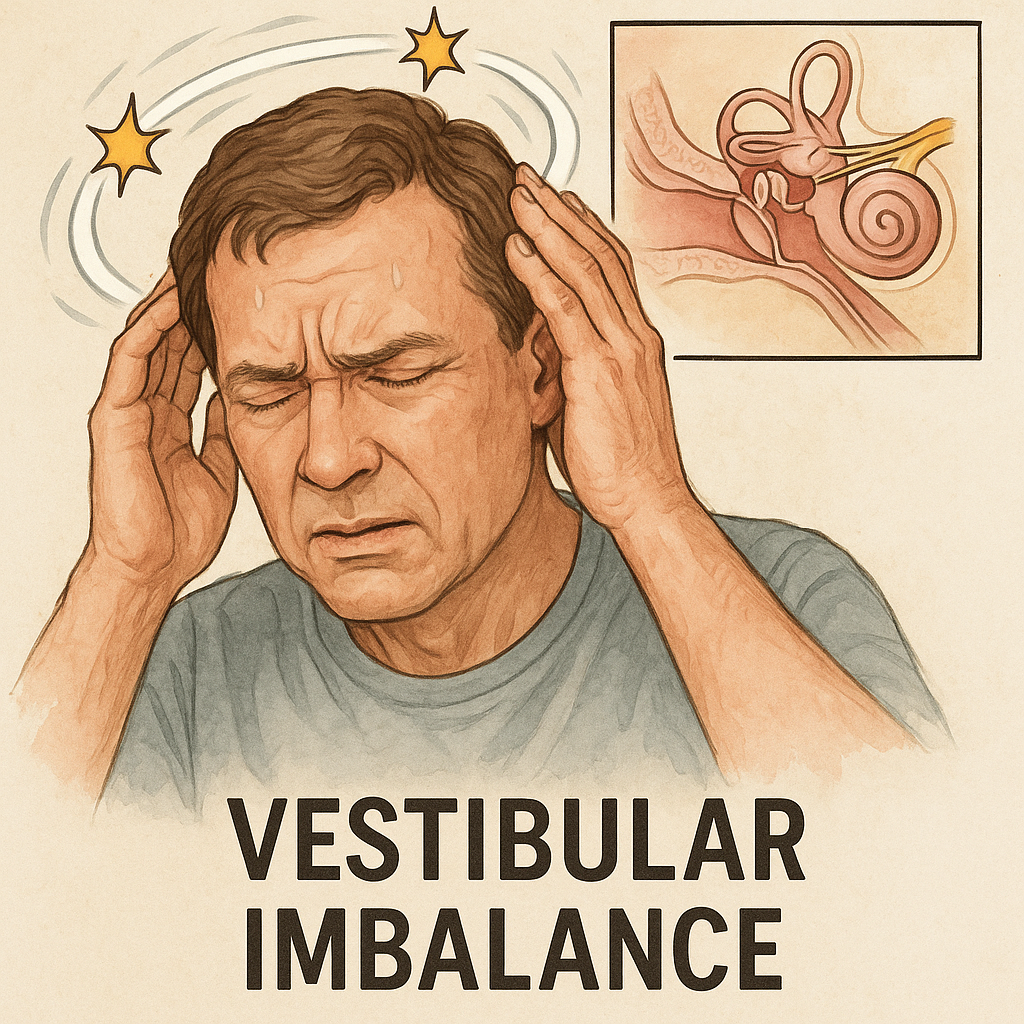
Benign Paroxysmal Positional Vertigo (BPPV)
What is it?
BPPV causes brief spinning sensations when you move your head, like turning in bed or looking up.
It happens when tiny crystals in the inner ear move out of place.
The spinning (vertigo) lasts seconds to a minute and then stops.
Hearing is not affected.
Though unpleasant, it is harmless and treatable.
What causes it?
The inner ear contains small crystals that help sense movement and balance.
When these crystals move into the wrong part of the ear canal, they disturb balance signals.
It can happen after a head injury, infection, or simply with age.
Sometimes it occurs without a clear reason.
Lying down or turning quickly can trigger episodes.
How is it diagnosed?
Doctors perform a simple head movement test called the Dix-Hallpike maneuver.
The test triggers the typical spinning sensation and specific eye movements.
Hearing and brain function tests are normal in BPPV.
Scans are rarely needed unless symptoms are unusual.
Diagnosis is based mainly on the pattern of dizziness.
What is the treatment?
Special head and body movements, like the Epley maneuver, reposition the crystals.
These are quick, noninvasive treatments done in the clinic.
Home exercises can help prevent recurrence.
Dizziness medicines are not usually needed.
Most people recover fully after one or two treatments.
What is the prognosis?
BPPV is very treatable and often resolves completely.
Some people may have mild recurrences over time.
Simple maneuvers can easily manage repeat episodes.
It does not cause permanent damage or hearing loss.
Avoiding prolonged bed rest can reduce the risk of recurrence.
Botulism
What is it?
Botulism is a rare but serious illness caused by a nerve toxin made by bacteria.
It leads to muscle weakness starting in the face and moving downward.
People stay awake and alert but become weak or paralyzed.
It can affect breathing and needs urgent hospital care.
With early treatment, most people recover fully.
What causes it?
The illness is caused by toxins from the bacteria Clostridium botulinum.
It can come from eating improperly canned or preserved food.
Babies can get it from swallowing spores that grow in their intestines.
It can also occur through wounds, especially in drug users.
The toxin blocks signals from nerves to muscles, causing paralysis.
How is it diagnosed?
Doctors look for typical signs — drooping eyelids, slurred speech, and descending weakness.
Tests can detect the toxin in blood, stool, or food samples.
Nerve tests show a pattern different from other nerve diseases.
Imaging or spinal tap may be done to rule out other causes.
Diagnosis is mainly clinical and treatment starts right away.
What is the treatment?
An antitoxin is given immediately to stop the toxin from doing more harm.
Hospital care supports breathing and swallowing if needed.
Patients may need a ventilator until muscle strength returns.
Antibiotics are used for wound infections.
Early treatment greatly improves survival and recovery.
What is the prognosis?
Recovery can take weeks to months as nerves slowly heal.
Most people recover completely with good medical care.
Early antitoxin use prevents complications.
Breathing problems are the main life-threatening risk.
Once recovered, the illness does not recur.
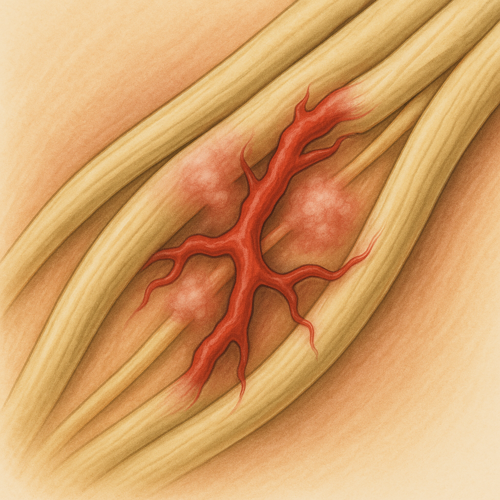
Brachial Plexopathy
What is it?
Brachial plexopathy is an injury to the network of nerves that controls the shoulder, arm, and hand.
It can cause weakness, numbness, or pain in one arm.
These nerves run from the neck to the arm and can be stretched, compressed, or inflamed.
The problem may affect part or all of the arm depending on the location.
Recovery varies depending on the cause and severity of the injury.
What causes it?
It can occur after trauma, such as a fall, accident, or surgery near the shoulder or chest.
Repetitive strain or radiation treatment can injure the nerve network.
Sometimes it’s caused by inflammation after infection (Parsonage-Turner syndrome).
Cancer pressing on the nerves may also cause it.
Rarely, it happens without a clear reason.
How is it diagnosed?
Doctors check muscle strength, sensation, and reflexes in the arm.
Nerve tests (EMG/NCS) help locate and measure the nerve injury.
MRI scans can show damage, swelling, or tumors affecting the nerves.
Blood tests may help find autoimmune or infection causes.
Diagnosis helps plan treatment and predict recovery.
What is the treatment?
Pain is managed with medicines or short courses of steroids.
Physical and occupational therapy help maintain movement and strength.
Severe injuries may need surgery or nerve grafting.
Underlying causes like tumors or infections are treated directly.
Patience and regular follow-up are important for recovery.
What is the prognosis?
Most people improve gradually over months.
Mild cases often recover fully.
Severe injuries or complete nerve tears may leave some weakness.
Recovery depends on the cause and speed of treatment.
Early therapy helps prevent stiffness and improves function.
Brain Abscess
What is it?
A brain abscess is a pocket of infection and pus inside the brain.
It happens when bacteria or fungi reach the brain and cause inflammation.
The swelling can increase pressure inside the skull.
It is a serious condition needing urgent treatment.
With modern care, most people recover well.
What causes it?
Infections from the ear, sinuses, or teeth can spread to the brain.
Germs can also travel through the bloodstream from other body infections.
Head injuries or surgeries can allow bacteria to enter.
People with weak immune systems are at higher risk.
Common bacteria include Streptococcus and Staphylococcus.
How is it diagnosed?
MRI or CT scans show a round area of infection with swelling.
Doctors look for fever, headache, and weakness on one side.
Blood tests and cultures help identify the germs.
Sometimes, a small needle procedure removes pus for testing.
Other brain conditions are ruled out using scans.
What is the treatment?
Strong intravenous antibiotics are started right away.
Surgery may be needed to drain large or deep abscesses.
Medicines are adjusted once test results identify the infection.
Seizure medicines are given to prevent convulsions.
Hospital monitoring ensures the swelling and infection resolve.
What is the prognosis?
Most patients recover fully with timely treatment.
Some may have mild lingering weakness or seizures.
Delay in treatment can cause serious complications.
Survival rates are now very high due to modern antibiotics.
Early diagnosis gives the best outcome.
Carpal Tunnel Syndrome
What is it?
Carpal tunnel syndrome happens when a nerve in your wrist, called the median nerve, gets squeezed.
This nerve controls feeling in your thumb, index, and middle fingers and helps move your hand.
When it’s under pressure, your hand may feel numb, tingly, or weak.
The symptoms often come on slowly and may feel worse at night.
It’s one of the most common nerve problems affecting the hand.
What causes it?
Repetitive hand and wrist movements—like typing or using tools—can irritate the nerve.
Health conditions such as diabetes, thyroid problems, or arthritis can increase the risk.
Fluid buildup in pregnancy can temporarily compress the nerve.
Injury or swelling in the wrist may make the space too tight for the nerve.
Sometimes, no single cause is found—it’s a combination of strain and anatomy.
How is it diagnosed?
Doctors ask about symptoms like tingling, numbness, and night pain.
A physical exam includes tapping or bending the wrist to reproduce symptoms.
Nerve tests (EMG or nerve conduction studies) check how signals travel through the wrist.
An ultrasound may show swelling around the nerve.
Blood tests might be done to rule out thyroid or sugar-related causes.
What is the treatment?
Wearing a wrist splint—especially at night—helps keep the wrist straight and relieves pressure.
Taking breaks from repetitive hand work is important.
Anti-inflammatory medicines or steroid injections can ease swelling and pain.
If severe, surgery can release pressure on the nerve and prevent long-term damage.
Gentle hand exercises help restore strength and movement after recovery.
What is the prognosis?
Most people get better with simple treatments and rest.
Early diagnosis leads to a quicker and more complete recovery.
If untreated, the hand can become weak and lose coordination.
Surgical results are usually excellent, especially when done early.
Avoiding repetitive strain helps prevent future problems.
Cauda Equina Syndrome
What is it?
Cauda equina syndrome is a serious condition where nerves at the bottom of the spine are squeezed.
These nerves control movement and sensation in the legs, bladder, and bowel.
It causes pain, numbness, and sometimes loss of bladder or bowel control.
It is a medical emergency and needs immediate treatment.
Without quick care, nerve damage can become permanent.
What causes it?
The most common cause is a large slipped disc in the lower back.
Other causes include spinal injuries, tumors, or severe infections.
Sometimes, bleeding or swelling after back surgery can compress the nerves.
In rare cases, inflammation or narrowing of the spinal canal is responsible.
Anything that takes up space in the spinal canal can trigger this condition.
How is it diagnosed?
Doctors look for red flag signs like leg weakness, saddle numbness, or bladder problems.
An MRI scan shows whether the nerves are being compressed.
Reflex tests and sensation checks help locate the affected area.
A bladder scan may show if urine is being retained.
The diagnosis must be made quickly to start urgent treatment.
What is the treatment?
Emergency surgery is needed to relieve pressure on the nerves.
The goal is to restore leg and bladder function as soon as possible.
Pain relief and antibiotics may be given if infection is involved.
A urinary catheter might be used to manage bladder issues.
After surgery, physiotherapy helps regain strength and mobility.
What is the prognosis?
Recovery depends on how quickly treatment begins—earlier is better.
Many patients regain walking ability and bladder control.
Some may have lasting numbness or weakness in the legs.
Long-term physiotherapy can help improve daily function.
Regular follow-up ensures that nerve recovery is monitored.
Cavernous Sinus Thrombosis
What is it?
Cavernous sinus thrombosis is a rare but serious blood clot near the base of the brain.
It often starts from an infection in the face, nose, or sinuses that spreads inward.
The condition can affect vision and eye movement and cause severe pain.
It needs urgent treatment to prevent brain or eye damage.
With modern antibiotics, it’s rare but still life-threatening if untreated.
What causes it?
Bacteria from sinus or facial infections can spread to the veins near the brain.
The veins in this area don’t have valves, allowing infection to move easily.
Common germs include Staphylococcus and Streptococcus species.
Less often, dental infections or ear infections are the source.
Rarely, trauma or surgery in the head region can trigger it.
How is it diagnosed?
Symptoms like fever, headache, eye swelling, and double vision raise suspicion.
An MRI scan with vein imaging confirms the clot.
Blood tests and cultures identify the infection.
Eye exams help check for pressure or nerve damage.
Doctors rule out other conditions like orbital cellulitis or aneurysm.
What is the treatment?
High-dose IV antibiotics are started right away to stop the infection.
Blood thinners may be used to prevent the clot from growing.
If a sinus infection or abscess is the source, it may need surgical drainage.
Pain relief, hydration, and careful monitoring are part of hospital care.
Treatment continues for several weeks to fully clear the infection.
What is the prognosis?
With early treatment, many patients recover well.
Vision or nerve problems may take time to improve.
Delay in care can lead to serious brain or eye complications.
Lifelong problems are uncommon but can occur in severe cases.
Timely antibiotic treatment saves lives and prevents recurrence.
Cerebral Aneurysm
What is it?
A cerebral aneurysm is a weak spot in a brain artery that bulges like a balloon.
If it leaks or bursts, it can cause sudden bleeding around the brain.
This is known as a subarachnoid hemorrhage and is a medical emergency.
Some aneurysms are small and found by chance before they rupture.
Prompt treatment can prevent bleeding and save lives.
What causes it?
High blood pressure and smoking weaken artery walls over time.
Some people inherit a tendency to develop aneurysms.
Conditions like polycystic kidney disease and connective tissue disorders can increase risk.
Head injury or infection may rarely cause one to form.
Aging and atherosclerosis make arteries more fragile.
How is it diagnosed?
A sudden, severe headache (“worst ever”) often points to a rupture.
A CT scan quickly detects bleeding around the brain.
Angiography shows the exact location and size of the aneurysm.
If the CT is normal but suspicion remains, a spinal tap checks for blood in spinal fluid.
Some aneurysms are found incidentally during imaging for other reasons.
What is the treatment?
Emergency care focuses on stopping bleeding and controlling blood pressure.
Surgeons may repair the aneurysm by clipping it or sealing it from inside using coiling.
Medications like nimodipine help prevent brain spasms after bleeding.
Fluid management and rest in a hospital’s ICU are part of recovery.
Lifestyle changes and follow-up imaging help prevent new aneurysms.
What is the prognosis?
Recovery depends on how quickly bleeding is controlled.
Many people recover fully if treated before or soon after rupture.
Delays can cause stroke, brain damage, or death.
After treatment, most patients live normal lives with routine checkups.
Avoiding smoking and managing blood pressure lowers recurrence risk.
Cerebral Palsy
What is it?
Cerebral palsy is a group of conditions that affect movement, posture, and muscle control.
It happens when a baby’s brain is injured or develops abnormally, often before or around the time of birth.
The condition is not progressive—meaning it doesn’t get worse over time—but symptoms can change as a child grows.
The severity can vary widely, from mild clumsiness to significant difficulty walking and moving.
It often affects balance, coordination, and fine motor skills.
What causes it?
The main cause is brain injury before, during, or shortly after birth.
Common factors include premature birth, lack of oxygen, infections during pregnancy, or brain bleeding.
Sometimes, no clear cause can be found even after detailed testing.
It is not caused by poor parenting or anything done after birth.
The brain damage does not worsen, but muscle stiffness or weakness may become more noticeable with growth.
How is it diagnosed?
Doctors usually diagnose cerebral palsy by observing a child’s movement and development over time.
A brain MRI may help find signs of past injury or abnormal development.
Developmental delays, stiffness, or unusual muscle tone often prompt early evaluation.
Other tests may be done to rule out genetic or metabolic conditions with similar symptoms.
Early diagnosis allows therapies to begin as soon as possible, improving outcomes.
What is the treatment?
Treatment focuses on helping the child move better and live as independently as possible.
Physical, occupational, and speech therapy are key parts of care.
Medications such as baclofen or botulinum toxin can reduce stiffness and spasms.
In severe cases, surgery may help improve posture or walking ability.
A team of specialists—therapists, doctors, and educators—works together for long-term support.
What is the prognosis?
Children with cerebral palsy can live full, meaningful lives with the right support and treatment.
Many learn to walk, communicate, and attend school with assistance.
The condition does not shorten life expectancy in most cases.
Early and ongoing therapy helps maximize function and confidence.
Regular follow-ups ensure adjustments to treatment as the child grows.
Cerebral Venous Sinus Thrombosis (CVST)
What is it?
CVST happens when a blood clot forms in the veins that drain blood from the brain.
This blockage can cause pressure to build up, leading to headaches, swelling, or even small strokes.
It’s a rare but serious cause of stroke, especially in young adults.
It affects the brain’s veins, not arteries, making it different from the usual type of stroke.
With timely treatment, most people recover fully.
What causes it?
Clots form due to conditions that make blood thicker or more likely to clot.
Common causes include dehydration, pregnancy, infections, or use of birth control pills.
Blood disorders such as factor V Leiden or antiphospholipid syndrome increase risk.
Sometimes, head injuries or certain cancers may also trigger clot formation.
In some cases, the exact cause is never found.
How is it diagnosed?
Doctors may suspect CVST when a person has severe or unusual headaches, vision changes, or seizures.
MRI or MR venography scans show the blocked veins and help confirm the diagnosis.
CT scans can also detect signs of clot or bleeding.
Blood tests may check for clotting disorders or other risk factors.
Eye exams can show swelling of the optic nerves caused by increased brain pressure.
What is the treatment?
The main treatment is blood-thinning medication to stop the clot from growing and allow healing.
Even if there is bleeding, anticoagulants are usually safe and necessary.
Pain relief and fluids help manage symptoms and prevent complications.
In severe cases, procedures to remove the clot or reduce pressure may be done.
Treating the cause—like stopping hormonal therapy or managing infections—is essential.
What is the prognosis?
Most people recover well, especially when diagnosed early.
Headaches and fatigue may last for some weeks but usually improve with time.
A few patients may have lasting weakness or vision problems if large areas were affected.
Recurrence is uncommon with proper treatment and follow-up.
Lifelong blood thinners are rarely needed unless an underlying clotting disorder is found.
Cervical Dystonia
What is it?
Cervical dystonia causes involuntary twisting or turning of the neck muscles.
The head may pull to one side, tilt forward or backward, or tremble slightly.
It can cause pain and stiffness in the neck and shoulders.
The condition is long-term but not dangerous or life-threatening.
It affects adults more often, particularly women in midlife.
What causes it?
It results from abnormal signals in the brain areas that control movement.
Most cases occur for no known reason (idiopathic), though some follow injury or certain drugs.
Family history may increase risk in some individuals.
Rarely, it occurs as part of another neurological condition.
Stress and fatigue can make the symptoms worse but do not cause the disease.
How is it diagnosed?
Diagnosis is based mainly on physical examination and medical history.
Doctors look for involuntary neck movements, abnormal postures, and muscle tightness.
Imaging tests like MRI help rule out other causes if symptoms appear suddenly.
Sometimes, nerve or muscle tests are used to guide treatment planning.
There is no single test to confirm it—clinical evaluation is key.
What is the treatment?
Botulinum toxin (Botox) injections into neck muscles are the most effective treatment.
Physical therapy helps keep muscles flexible and reduce pain.
Medicines like muscle relaxants or anticholinergics may ease symptoms.
Deep brain stimulation surgery may be considered if other treatments fail.
Relaxation, stress management, and good posture can help daily control.
What is the prognosis?
Many people see good relief with Botox injections and therapy.
Symptoms may fluctuate but often stabilize over time.
The condition usually does not affect life expectancy.
Regular follow-up helps adjust treatments as needed.
Early intervention improves comfort and quality of life.
Cervical Myelopathy
What is it?
Cervical myelopathy is a condition where the spinal cord in the neck becomes squeezed or compressed.
This pressure affects the signals that travel between the brain and the rest of the body.
It can cause problems with balance, walking, hand coordination, and bladder control.
The condition usually develops slowly over time as the spine ages or wears down.
It is one of the most common causes of spinal cord problems in older adults.
What causes it?
The most common cause is wear-and-tear arthritis in the neck, known as cervical spondylosis.
Bone spurs, thickened ligaments, or bulging discs can press on the spinal cord.
Injuries or previous spine surgeries may also lead to narrowing of the spinal canal.
Some people are born with a smaller spinal canal, making them more prone to compression.
Repeated stress or movement of the neck over the years can worsen the problem.
How is it diagnosed?
Doctors diagnose it by checking for weakness, stiffness, or coordination problems in the arms and legs.
MRI scans show whether the spinal cord is being compressed and where.
X-rays or CT scans may reveal bone changes or misalignment.
Sometimes nerve tests help rule out other conditions like neuropathy or multiple sclerosis.
Early diagnosis is important because symptoms can worsen if untreated.
What is the treatment?
Mild cases may improve with physical therapy, gentle exercise, and neck support.
Medications can help reduce pain and muscle stiffness.
Surgery is often needed when compression is severe or symptoms are progressing.
Operations relieve pressure by removing bone spurs or discs and stabilizing the spine.
After surgery, rehabilitation helps regain balance, strength, and confidence.
What is the prognosis?
Many people improve or stabilize after treatment, especially with early surgery.
Long-standing compression may leave some residual stiffness or numbness.
Without treatment, symptoms can slowly worsen and cause permanent nerve damage.
Recovery depends on the extent of spinal cord injury before surgery.
Regular follow-ups ensure ongoing spinal health and prevent recurrence.
Charcot-Marie-Tooth Disease
What is it?
Charcot-Marie-Tooth (CMT) disease is a group of inherited conditions that damage the nerves controlling movement and sensation.
It mainly affects the feet and legs first, then sometimes the hands.
People often notice weakness, balance issues, or high-arched feet.
Symptoms develop slowly over years and vary in severity.
Despite the name, it has nothing to do with the teeth—it’s named after the doctors who discovered it.
What causes it?
CMT is caused by changes (mutations) in genes that affect the nerves in the arms and legs.
These genetic changes can run in families, though some cases occur spontaneously.
The faulty genes damage either the nerve fibers or the insulating myelin covering them.
This makes it harder for electrical signals to travel between the brain and muscles.
Different types of CMT are linked to different genes but cause similar symptoms.
How is it diagnosed?
Doctors look for signs like muscle weakness, high arches, or absent reflexes.
Nerve tests (electromyography and nerve conduction studies) show how fast signals travel.
Genetic testing confirms the exact type of CMT and whether it runs in the family.
Family history helps identify inherited patterns.
Occasionally, a nerve biopsy is done to study nerve structure directly.
What is the treatment?
There is no cure, but treatment focuses on managing symptoms and maintaining function.
Physical and occupational therapy help strengthen muscles and improve mobility.
Braces and special shoes support weak ankles and prevent tripping.
Surgery can correct severe foot deformities in some cases.
Counseling and support groups help families cope with long-term challenges.
What is the prognosis?
Most people with CMT lead normal lives with good symptom management.
The condition progresses slowly and does not usually shorten life expectancy.
Regular exercise and therapy can help maintain strength and prevent falls.
Mobility aids or braces may be needed over time, depending on severity.
Ongoing medical care ensures comfort and independence through all stages.
Chronic Inflammatory Demyelinating Polyneuropathy (CIDP)
What is it?
CIDP is a rare nerve disorder that causes weakness, numbness, and loss of reflexes.
It happens when the immune system mistakenly attacks the protective covering (myelin) of the peripheral nerves.
This slows down nerve signals and makes muscles weaker.
CIDP develops gradually over weeks or months, unlike sudden-onset Guillain-Barré syndrome.
It affects both sides of the body, usually starting in the legs.
What causes it?
CIDP is an autoimmune condition, meaning the body’s defense system targets its own nerves.
The exact trigger is unknown, but infections or immune reactions may play a role.
Some cases are linked to diabetes, HIV, or certain blood disorders.
It is not contagious or inherited.
The ongoing inflammation damages the myelin sheath, leading to long-term nerve issues if untreated.
How is it diagnosed?
Diagnosis is based on medical history, symptoms, and neurological examination.
Nerve tests show slowed signal conduction due to demyelination.
A lumbar puncture (spinal tap) may show high protein levels in the spinal fluid.
MRI scans can reveal thickened nerve roots or inflammation.
Doctors rule out other causes like diabetes or genetic neuropathies before confirming CIDP.
What is the treatment?
Treatment calms the immune system and helps nerves recover.
Corticosteroids, IV immunoglobulin (IVIG), or plasma exchange are the main treatments.
Some patients may need long-term medication to prevent relapses.
Physical therapy helps rebuild strength and coordination.
Regular monitoring ensures that treatment remains effective over time.
What is the prognosis?
Most people improve significantly with proper treatment.
Some recover fully, while others may have mild lingering weakness.
Without treatment, symptoms can worsen and cause permanent nerve damage.
Relapses can occur but are usually manageable with ongoing care
CLIPPERS (Chronic Lymphocytic Inflammation with Pontine Perivascular Enhancement Responsive to Steroids)
What is it?
CLIPPERS is a rare inflammation that affects the brainstem—the area that controls balance, speech, and eye movements.
The inflammation causes small areas of swelling that can be seen on an MRI scan.
It responds very well to steroid medicines, which reduce inflammation and symptoms.
The condition is not infectious or hereditary.
With proper treatment, most people recover well and return to normal activity.
What causes it?
The exact cause is not known, but it is thought to be an autoimmune condition, where the body’s immune system attacks healthy tissue.
It is not caused by infection, injury, or cancer.
Some researchers believe it may be related to other inflammatory diseases of the brain.
It is not linked to lifestyle or environmental factors.
There is no known genetic cause.
How is it diagnosed?
MRI scans of the brain show small, bright, “pepper-like” spots in the brainstem.
A spinal tap (lumbar puncture) may show mild inflammation in the fluid around the brain.
Blood tests help rule out infections or other autoimmune conditions.
Occasionally, a brain biopsy is needed if the diagnosis is uncertain.
The symptoms improve rapidly with steroids, which helps confirm the diagnosis.
What is the treatment?
Steroid medications such as prednisone or methylprednisolone are the main treatment.
After symptoms improve, a lower steroid dose or other medicines may be used to prevent relapse.
Regular MRI scans help monitor recovery.
Physical therapy can help with balance and coordination problems.
Most patients respond quickly and dramatically to treatment.
What is the prognosis?
Most people recover well with treatment.
Some may need long-term low-dose therapy to prevent relapse.
Ongoing medical follow-up ensures early treatment if symptoms return.
The condition does not usually lead to permanent disability if treated early.
Quality of life is generally good with proper care.
Cluster Headache
What is it?
Cluster headache causes severe, one-sided pain around the eye or temple.
Attacks come in “clusters,” meaning several headaches a day for weeks or months, followed by pain-free periods.
Each attack typically lasts 15 to 180 minutes.
The pain is often described as sharp, burning, or piercing.
It’s sometimes called the “suicide headache” because of its intensity, but treatment is available and effective.
What causes it?
The exact cause is not known, but it involves overactivity in a part of the brain called the hypothalamus.
Triggers can include alcohol, strong smells, or changes in sleep pattern.
It is more common in men and often starts between ages 20 and 50.
Smoking increases the risk and can make attacks worse.
It is not hereditary, though it can run in some families.
How is it diagnosed?
Diagnosis is based on the pattern of attacks and symptoms.
Pain is always on one side, often with tearing, nasal stuffiness, or redness of the eye.
MRI scans may be done to rule out other causes of one-sided headache.
Keeping a headache diary helps track timing and triggers.
Doctors may refer to a neurologist for confirmation.
What is the treatment?
Inhaled oxygen through a face mask can stop an attack quickly.
Medicines like sumatriptan (injection or nasal spray) also work fast to stop pain.
Preventive treatments such as verapamil or steroids reduce attack frequency.
Avoiding alcohol during active cluster periods can prevent triggering an attack.
In rare cases, nerve stimulation treatments may be considered if standard therapy fails.
What is the prognosis?
Cluster periods may recur once or twice a year but often lessen over time.
With the right treatment plan, most people control their attacks successfully.
Lifestyle adjustments, stress management, and sleep regulation can help.
The condition is not life-threatening, though it can be very painful.
Regular follow-up with a neurologist helps adjust long-term therapy.
Complex Regional Pain Syndrome (CRPS)
What is it?
CRPS is a chronic pain condition that usually affects one arm or leg after an injury or surgery.
The pain is much more severe than expected from the original injury.
The affected area may feel burning, swollen, or extremely sensitive to touch.
Skin color or temperature may change, and movement may become difficult.
Early treatment gives the best chance for recovery.
What causes it?
CRPS happens when the nerves controlling pain and blood flow become overactive after injury.
Even a minor injury, fracture, or surgery can trigger it.
It is not psychological, though stress can worsen symptoms.
Some people may have a genetic tendency toward abnormal nerve responses.
The exact cause is not always clear.
How is it diagnosed?
Diagnosis is based on symptoms and physical examination.
Common findings include swelling, temperature changes, color changes, or unusual sweating.
Tests like X-rays, bone scans, or nerve studies may support the diagnosis.
There is no single test that confirms CRPS—clinical evaluation is key.
Early referral to a pain specialist or neurologist is important.
What is the treatment?
Physical and occupational therapy are vital to keep the limb moving and prevent stiffness.
Medications such as pain relievers, nerve pain drugs, or steroids may help.
Nerve blocks or spinal cord stimulation can reduce pain in severe cases.
Counseling and relaxation therapies help manage stress and coping.
A combination of treatments gives the best results.
What is the prognosis?
Recovery varies—some people improve completely, while others have long-term symptoms.
Early diagnosis and treatment improve outcomes significantly.
Keeping the limb active and following therapy plans are key.
Pain may lessen over time, even if mild symptoms remain.
Support groups and ongoing care help maintain function and quality of life.
Critical Illness Myopathy (CIM)
What is the condition?
Critical Illness Myopathy is a severe muscle weakness seen in patients who are critically ill and spend long periods in the ICU.
It mainly affects the shoulders, arms, and thighs, making it difficult to move or breathe independently.
It often appears after days or weeks of mechanical ventilation or severe infection.
Sensation remains normal, distinguishing it from nerve disorders.
It is one of the main causes of prolonged weakness after critical illness.
What causes the condition?
Prolonged bed rest and severe illness lead to muscle fiber damage and wasting.
High doses of steroids or muscle relaxants can worsen the weakness.
Inflammation and poor blood flow in critical illness further harm muscle tissue.
Low nutrition and lack of physical activity slow recovery.
The condition often develops alongside critical illness neuropathy.
How is it diagnosed?
Doctors suspect CIM when a patient stays weak even after the main illness improves.
Nerve and muscle tests (EMG) help confirm that weakness comes from the muscles.
Blood tests may show slightly raised muscle enzymes.
Muscle biopsy is sometimes done to confirm muscle damage.
Other causes of weakness, such as nerve or spinal problems, are ruled out.
How is it treated?
The focus is on treating the main illness and supporting recovery.
Reducing steroid use and avoiding unnecessary sedatives help prevent worsening.
Early physical therapy and mobilization strengthen weak muscles.
Proper nutrition and protein intake are important for healing.
Recovery may take several weeks to months, depending on severity.
What is the prognosis?
Most people recover gradually with good care and rehabilitation.
Some may have mild lingering weakness or fatigue for a few months.
Early therapy improves long-term outcomes.
Emotional and family support play a key role in recovery.
Overall prognosis is good if complications are prevented.
Critical Illness Neuropathy (CIN)
What is the condition?
Critical Illness Neuropathy is a nerve disorder seen in severely ill ICU patients.
It causes weakness in both arms and legs, often making it hard to breathe without support.
It affects the peripheral nerves that carry messages between brain and muscles.
Sensory symptoms like numbness or tingling may occur but are mild.
It commonly appears after severe infections or organ failure.
What causes the condition?
Severe infections and inflammation damage the nerves.
Low oxygen and poor blood flow worsen nerve injury.
High blood sugar and certain medications increase the risk.
Prolonged immobilization adds to weakness.
It may occur along with muscle damage (polyneuromyopathy).
How is it diagnosed?
Doctors notice persistent weakness even after the critical illness improves.
Nerve conduction tests show slow or absent nerve signals.
EMG helps distinguish nerve damage from muscle weakness.
Other causes like Guillain–Barré syndrome are excluded.
Diagnosis is clinical and supported by electrodiagnostic findings.
How is it treated?
There is no specific medicine; treatment is mainly supportive.
Managing infection, blood sugar, and nutrition helps recovery.
Early physical and occupational therapy prevent complications.
Doctors reduce harmful medications when possible.
Gradual mobilization improves nerve recovery and strength.
What is the prognosis?
Recovery usually begins once the underlying illness resolves.
Most people improve over weeks to months with therapy.
Severe cases may have lingering weakness for longer periods.
Early rehabilitation speeds up functional recovery.
With proper care, many regain near-normal strength.
Cubital Tunnel Syndrome
What is the condition?
Cubital Tunnel Syndrome occurs when the ulnar nerve is compressed behind the elbow.
This nerve controls feeling in the ring and little fingers and helps hand movement.
Pressure or stretching of the nerve causes numbness or tingling in these fingers.
Weakness and clumsiness in the hand may develop over time.
It is the second most common nerve entrapment after carpal tunnel syndrome.
What causes the condition?
Repeated elbow bending or leaning on the elbow can irritate the nerve.
Injury, arthritis, or thickened tissue may narrow the cubital tunnel.
Sleeping with bent elbows can increase pressure on the nerve.
Cysts or bone spurs may rarely compress the nerve.
Sometimes no single cause is identified.
How is it diagnosed?
A doctor checks sensation and strength in the affected hand.
Tapping the nerve at the elbow may reproduce tingling (Tinel’s sign).
Nerve conduction tests confirm slowing across the elbow.
Ultrasound or MRI may be used if another cause is suspected.
Early diagnosis prevents permanent nerve damage.
How is it treated?
Avoiding prolonged elbow flexion or pressure helps mild cases.
Night splints can keep the elbow straight and reduce symptoms.
Physiotherapy improves flexibility and hand strength.
Persistent or severe cases may need surgery to release the nerve.
Most people recover fully after treatment with proper care.
What is the prognosis?
Early treatment leads to excellent recovery.
Mild cases often resolve with rest and splints.
Surgery provides good long-term outcomes in most patients.
Untreated cases can cause muscle wasting and weakness.
With proper management, normal hand function usually returns.
Dermatomyositis
What is Dermatomyositis?
Dermatomyositis is a rare condition where the immune system attacks the muscles and skin.
It causes muscle weakness, especially in the shoulders and hips.
A red or purple rash often appears on the eyelids or knuckles.
Both adults and children can be affected.
Early treatment helps prevent long-term disability.
What causes it?
The immune system mistakenly targets blood vessels in muscle and skin.
This leads to inflammation and reduced blood supply to muscles.
Certain antibodies in the blood may play a role.
In adults, it may sometimes be linked to an underlying cancer.
The exact cause is not always known.
How is it diagnosed?
Blood tests show high muscle enzymes like creatine kinase (CK).
EMG can detect electrical changes in affected muscles.
Muscle biopsy confirms inflammation and damage.
Antibody tests may help identify specific subtypes.
Screening for hidden cancers is recommended in adults.
How is it treated?
Corticosteroids are the main treatment to reduce inflammation.
Other medicines like methotrexate or azathioprine may be added.
IV immunoglobulin (IVIG) is used for severe or resistant cases.
Physical therapy helps restore strength and mobility.
Treating any related cancer often improves the condition.
What is the prognosis?
Most people improve with early and consistent treatment.
Muscle strength may take months to fully recover.
Some patients experience relapses that need re-treatment.
Regular follow-up helps prevent complications.
With good care, many return to normal daily activities.
Diabetic Amyotrophy
What is Diabetic Amyotrophy?
Diabetic amyotrophy is a nerve condition linked to diabetes.
It causes severe pain and weakness in one thigh or hip.
The muscles may shrink and walking becomes difficult.
It mainly affects older adults with type 2 diabetes.
Recovery happens slowly over months to years.
What causes it?
High blood sugar damages small blood vessels that feed the nerves.
Inflammation reduces blood flow to the lumbosacral plexus.
Nerves controlling thigh and hip muscles become weak.
The process is partly immune-mediated.
Poor diabetes control increases the risk.
How is it diagnosed?
Diagnosis is based on pain and weakness in one leg.
EMG and nerve studies show nerve damage in the thigh muscles.
MRI may show inflammation in the nerve roots.
Blood tests rule out other causes of neuropathy.
A neurologist confirms the diagnosis after detailed testing.
How is it treated?
Strict blood sugar control helps prevent further nerve injury.
Pain is treated with medicines like gabapentin or duloxetine.
Physical therapy improves strength and balance.
In severe cases, steroids or IVIG may be used.
Supportive care and reassurance are very important.
What is the prognosis?
The condition usually improves over time.
Recovery may take several months to two years.
Pain and weakness lessen as nerves heal.
Most people regain independence in daily activities.
Good diabetes control reduces recurrence risk.
Diabetic Neuropathy
What is Diabetic Neuropathy?
Diabetic neuropathy is nerve damage caused by long-term high blood sugar.
It affects sensation, movement, and sometimes internal organs.
The most common form causes numbness and burning pain in the feet.
Over time, it may spread to the hands.
Good sugar control can help slow its progress.
What causes it?
High blood sugar injures small blood vessels that supply nerves.
This leads to poor oxygen flow and nerve damage.
Long-standing diabetes, high cholesterol, and smoking increase risk.
The damage builds up slowly over years.
Poor blood flow and inflammation make it worse.
How is it diagnosed?
Doctors check for numbness, pain, and loss of reflexes.
Tests may include nerve conduction studies or EMG.
Blood tests rule out vitamin or thyroid problems.
Foot exams look for ulcers or injuries from loss of feeling.
Early diagnosis helps prevent serious complications.
How is it treated?
The most important step is controlling blood sugar levels.
Pain can be managed with gabapentin, duloxetine, or pregabalin.
Regular foot care prevents sores and infections.
Exercise improves circulation and balance.
Stopping smoking and healthy diet support nerve healing.
What is the prognosis?
Symptoms may improve with better sugar control.
Nerve damage may not fully reverse but can stabilize.
Regular care helps prevent ulcers and amputations.
Early treatment gives the best outcome.
Most people lead active, independent lives with good care.
Essential Tremor
What is it?
Essential tremor causes rhythmic shaking, usually of the hands when performing actions.
It can also affect the head, voice, or legs in some people.
The tremor happens during movement, not at rest.
It often runs in families and can start at any age.
Though not dangerous, it can interfere with daily activities.
What causes it?
It results from overactive brain circuits that control movement.
The cerebellum, which coordinates movement, may be involved.
Genetics play a major role, especially when family members are affected.
Stress, caffeine, or tiredness can make tremor worse.
Small amounts of alcohol may briefly reduce shaking.
How is it diagnosed?
Doctors identify it by examining the tremor during movement.
It is diagnosed after ruling out other conditions like Parkinson’s disease.
Blood tests and brain scans may be done to exclude other causes.
The tremor usually disappears when the hands are at rest.
A family history of tremor helps confirm the diagnosis.
How is it treated?
Mild cases may not need treatment.
Medications such as propranolol or primidone can reduce tremor.
Physical therapy and weighted utensils can help with daily tasks.
Botulinum toxin injections may help with head or voice tremors.
In severe cases, deep brain stimulation can provide relief.
What is the prognosis?
The tremor may slowly worsen over time but remains manageable.
Many people lead normal, active lives with treatment.
Symptoms can vary day to day depending on stress or fatigue.
Most find medication or lifestyle adjustments helpful.
It does not shorten life expectancy.
Epilepsy
What is it?
Epilepsy is a condition where people have repeated seizures from bursts of brain activity.
Seizures can cause shaking, loss of awareness, or brief staring spells.
It can begin at any age but is most common in children and older adults.
Some people have one seizure type; others have several.
With proper care, most people live normal lives.
What causes it?
It can be caused by brain injury, stroke, infection, or birth problems.
Some people inherit a genetic tendency toward seizures.
In many cases, no clear cause is found.
The brain’s electrical signals become overactive and unbalanced.
Triggers like missed medication, stress, or flashing lights can cause seizures.
How is it diagnosed?
Diagnosis is based on a detailed seizure history.
EEG tests show abnormal brain electrical activity.
MRI scans may reveal scars or structural problems in the brain.
Blood tests can help rule out infections or metabolic causes.
Doctors use seizure type to choose the best treatment.
How is it treated?
Most people respond well to anti-seizure medications.
If two medicines fail, surgery or nerve stimulation may be options.
Getting enough sleep and avoiding alcohol helps reduce seizures.
Taking medications on time is very important.
Support and education help people live safely and confidently.
What is the prognosis?
Around 70% of people gain good control with medication.
Some may eventually stop medication after being seizure-free for years.
A few have drug-resistant epilepsy needing specialist care.
Most people can work, study, and drive safely once controlled.
With regular follow-up, long-term outlook is excellent.
Fabry’s Disease
What is it?
Fabry’s disease is a rare inherited condition that affects how the body breaks down certain fats.
These fats build up in blood vessels and organs like the kidneys, heart, and nerves.
It mainly affects men but can also appear in women with milder symptoms.
Common signs include burning pain in hands and feet, rashes, and heat intolerance.
Early diagnosis helps prevent long-term organ damage.
What causes it?
It is caused by a change in a gene that makes an enzyme called alpha-galactosidase A.
Without this enzyme, fatty substances build up inside body cells.
This buildup damages blood vessels and vital organs over time.
The condition is passed from parents to children through the X chromosome.
The amount of enzyme activity often determines how severe the disease becomes.
How is it diagnosed?
Doctors check enzyme levels in the blood, which are very low in affected males.
Genetic testing confirms the diagnosis and helps identify carriers.
Eye exams and skin findings can also suggest the disease.
Kidney and heart tests check for complications.
Family screening is important to find other affected relatives.
How is it treated?
Enzyme replacement therapy helps slow or stop disease progression.
Some patients may take oral medication if their mutation allows it.
Pain and nerve symptoms can be managed with special medicines.
Early treatment protects the kidneys, heart, and brain from damage.
Regular follow-up helps track progress and manage symptoms.
What is the outlook?
With early and ongoing treatment, many people live active lives.
Kidney and heart care improve long-term health.
Family testing helps prevent late diagnosis in others.
New research may bring more treatment options in the future.
Lifelong care with a specialist team is recommended.
Facioscapulohumeral Muscular Dystrophy (FSHD)
What is it?
FSHD is a muscle disease that causes slow, progressive weakness.
It mainly affects the face, shoulders, and upper arms.
People may have trouble closing their eyes, smiling, or lifting their arms.
It runs in families and can vary in severity.
Most people remain active and mobile for many years.
What causes it?
It occurs due to a genetic change that affects how certain muscle genes are turned on.
This leads to gradual muscle damage and weakness.
The condition is inherited in an autosomal dominant way.
Sometimes it appears for the first time in a family due to a new mutation.
Both men and women can be affected equally.
How is it diagnosed?
Diagnosis is based on muscle weakness pattern and family history.
Blood tests may show mildly raised muscle enzymes.
Genetic testing confirms the diagnosis.
Muscle biopsy or nerve tests may help rule out other causes.
Genetic counseling is offered to affected families.
How is it treated?
There is no cure, but supportive care helps maintain function.
Physical therapy and stretching prevent stiffness and contractures.
Braces can improve walking and posture.
Surgery may help fix shoulder blade weakness in some cases.
Regular follow-up ensures early detection of complications.
What is the outlook?
Most people remain able to walk and work independently.
The disease progresses slowly over many years.
Life expectancy is usually normal.
Emotional and family support help maintain quality of life.
Research is ongoing to develop targeted treatments.
Femoral Neuropathy
What is it?
Femoral neuropathy happens when the femoral nerve in the thigh is damaged.
It causes weakness in the front of the thigh and knee.
Some people notice numbness or tingling in the inner leg.
It can occur after surgery, injury, or due to bleeding inside the abdomen.
Early recognition helps recovery.
What causes it?
Damage can happen from surgery, trauma, or prolonged pressure on the nerve.
Diabetes and internal bleeding can also injure the nerve.
The nerve may be stretched or compressed during pelvic or hip procedures.
Infections or tumors pressing on the nerve are rare causes.
Identifying the cause is key for proper treatment.
How is it diagnosed?
Doctors test muscle strength and sensation in the leg.
Reflex testing shows loss of the knee-jerk reflex.
Nerve and muscle tests confirm the diagnosis.
Imaging may be done to look for bleeding or pressure on the nerve.
Other conditions like spinal problems may need to be ruled out.
How is it treated?
Treating the underlying cause is the first step.
Physical therapy strengthens muscles and prevents stiffness.
Knee braces may help with walking and stability.
Pain medicines like gabapentin or duloxetine can help nerve pain.
Most people improve over time with care and therapy.
What is the outlook?
Recovery depends on the cause and severity of nerve injury.
Surgical injuries often improve within months.
Diabetes-related cases may take longer to heal.
Ongoing physiotherapy improves strength and mobility.
Most patients regain good leg function with treatment.
Fragile X Syndrome
What is it?
Fragile X syndrome is a genetic condition causing learning and behavior difficulties.
It is the most common inherited cause of intellectual disability.
Boys are usually more affected than girls.
Many children show features of autism such as poor eye contact or hand flapping.
Early support helps improve learning and social skills.
What causes it?
It happens due to a change in the FMR1 gene on the X chromosome.
The gene fails to make enough of a protein needed for brain development.
This affects communication between brain cells.
The condition is inherited from a parent who carries the abnormal gene.
The defect can get worse over generations.
How is it diagnosed?
Genetic testing confirms the diagnosis by identifying the FMR1 gene change.
Blood tests are simple and accurate for detection.
Doctors may recommend testing other family members.
Early diagnosis allows early developmental therapies.
Brain scans or EEGs may be used if seizures occur.
How is it treated?
There is no cure, but therapy helps improve communication and behavior.
Speech and occupational therapy support learning and social skills.
Medicines can help manage anxiety, hyperactivity, or seizures.
Behavioral therapy helps parents and teachers support the child.
Regular medical and developmental follow-up is important.
What is the outlook?
Many children learn to communicate and live semi-independently.
Girls often have milder symptoms than boys.
Lifelong support may be needed for some individuals.
Family counseling helps with understanding inheritance risks.
Research continues toward new targeted treatments.
Friedreich’s Ataxia
What is it?
Friedreich’s ataxia is a rare inherited disorder affecting balance and coordination.
It usually begins in childhood or the teenage years.
People may develop difficulty walking, slurred speech, and weakness.
It affects both the nervous system and the heart.
The disease progresses slowly over time.
What causes it?
It is caused by a change in the FXN gene, which controls energy production in cells.
The defect leads to low levels of a protein called frataxin.
Nerve cells and heart muscle are most affected.
The condition is passed down from both parents (recessive inheritance).
Damage comes from buildup of iron and stress inside cells.
How is it diagnosed?
Genetic testing confirms the diagnosis.
Doctors may do nerve tests and MRI scans to check the spine and brain.
Heart and blood sugar tests are important to detect complications.
Family members may also need genetic counseling.
Early detection allows supportive therapies to begin sooner.
How is it treated?
There is no cure, but therapy helps maintain movement and independence.
Physical therapy, braces, and mobility aids improve walking.
Heart and diabetes treatments manage related complications.
Surgery may be needed for spine or foot deformities.
Research on gene therapy and antioxidants is ongoing.
What is the outlook?
The condition progresses gradually over years.
Many patients need wheelchairs by young adulthood.
With good medical care, heart and diabetes issues can be managed.
Supportive therapy improves quality of life.
New treatments may change the future outlook.
Frontotemporal Dementia (FTD)
What is it?
FTD is a brain disorder that causes personality, behavior, and language changes.
It often begins between ages 45 and 65.
Memory is usually preserved early, unlike in Alzheimer’s disease.
It can make people act impulsively or lose interest in daily activities.
Families often notice emotional and social changes first.
What causes it?
FTD happens when nerve cells in the front and side parts of the brain die.
Abnormal proteins build up inside brain cells, leading to damage.
Some cases are inherited from parents due to genetic mutations.
Stress and aging may speed up changes in brain function.
The disease slowly worsens over several years.
How is it diagnosed?
Doctors evaluate behavior, speech, and thinking changes.
Brain scans show shrinkage in the front and temporal lobes.
Memory tests help distinguish it from other dementias.
Family history and genetic testing may guide diagnosis.
Early recognition allows better planning and support.
How is it treated?
There is no cure, but treatments help manage symptoms.
Medicines can reduce anxiety, agitation, or impulsive behavior.
Speech therapy supports communication for language problems.
Counseling and caregiver support are essential for families.
Regular follow-up ensures safety and emotional wellbeing.
What is the outlook?
FTD progresses over several years, with increasing dependence.
Emotional and behavioral symptoms can be distressing for families.
Early planning helps manage care needs effectively.
Support groups and counseling improve coping and quality of life.
Research on new drugs and therapies offers future hope.
Genitofemoral Neuralgia
What is it?
Genitofemoral neuralgia is a painful condition affecting the groin and genital area.
It happens when the genitofemoral nerve is irritated or injured.
The pain is usually burning or sharp and may spread to the thigh.
It can make walking, standing, or sexual activity uncomfortable.
Although not dangerous, it can cause significant distress and limit movement.
What causes it?
It often occurs after abdominal or groin surgery, especially hernia repair.
The nerve may be stretched, cut, or trapped in scar tissue.
Less commonly, it may follow trauma, infection, or tumors pressing on the nerve.
The nerve irritation leads to overactive pain signals.
In some cases, no clear cause is found.
How is it diagnosed?
Doctors identify it by the location and type of pain.
The pain usually worsens with standing and eases when sitting or bending the hip.
A local anesthetic injection that relieves the pain helps confirm the diagnosis.
Imaging such as MRI may rule out other causes of pain.
It is important to distinguish it from other nearby nerve problems.
How is it treated?
Mild cases may improve with rest, heat, and gentle movement.
Medicines for nerve pain such as gabapentin or duloxetine may help.
Nerve blocks or minor procedures can relieve pain for weeks or months.
Surgery is rarely needed and reserved for severe, persistent pain.
Supportive therapy and relaxation techniques improve daily comfort.
What is the prognosis?
Many people recover with medication and simple treatments.
Early recognition helps avoid chronic pain.
Nerve pain may come and go but usually becomes milder over time.
A small number may have persistent symptoms needing long-term care.
Most patients can return to normal activity with proper management.
Glossopharyngeal Neuralgia
What is it?
Glossopharyngeal neuralgia causes brief, severe pain in the throat, tongue, or ear.
It affects the glossopharyngeal nerve, which helps control swallowing and sensation in the throat.
The pain feels like an electric shock lasting a few seconds.
Episodes may happen many times a day, triggered by talking or swallowing.
Though painful, it does not cause lasting nerve damage.
What causes it?
It often occurs when a blood vessel presses on the glossopharyngeal nerve.
The nerve may also be affected by inflammation or scarring.
Rarely, a tumor or multiple sclerosis can irritate the nerve.
Sometimes no specific cause is found.
The result is sudden bursts of pain from overactive nerve signals.
How is it diagnosed?
Doctors diagnose it based on the pattern and triggers of the pain.
The pain is usually one-sided and located deep in the throat or ear.
MRI scans may check for blood vessel pressure or other causes.
A numbing medicine applied to the throat that stops the pain confirms the diagnosis.
Other causes of throat or ear pain must be ruled out.
How is it treated?
Medications such as carbamazepine or gabapentin reduce nerve overactivity.
Local anesthetic injections can provide temporary relief.
Surgery may be needed if medicines stop working.
Microvascular decompression relieves pressure on the nerve and often cures the pain.
Relaxation and stress management help reduce pain triggers.
What is the prognosis?
Most people respond well to medication.
Some may need long-term treatment to prevent recurrences.
Surgery can provide lasting relief in severe cases.
The condition is not life-threatening but can impact quality of life.
Regular follow-up helps adjust therapy and maintain comfort.
HaNDL Syndrome
What is it?
HaNDL syndrome is a rare condition that causes short-lasting headaches and temporary neurological symptoms like weakness or speech difficulty.
These symptoms can resemble a stroke but always recover completely.
The condition often appears suddenly and may last for a few weeks to months.
It is not dangerous and usually goes away on its own.
Most people make a full recovery without long-term problems.
What causes it?
The exact cause is unknown, but it may follow a mild viral infection.
It is thought to be related to temporary inflammation in the brain’s coverings (meninges).
This inflammation may briefly disturb blood flow or nerve function.
It is not inherited or caused by stress.
The condition is self-limiting and rarely comes back after recovery.
How is it diagnosed?
Diagnosis is made by recognizing the pattern of symptoms and ruling out other serious causes.
A brain scan (MRI) is usually normal.
A lumbar puncture may show a few extra white cells, suggesting mild inflammation.
Blood tests and scans help rule out infection or stroke.
A neurologist usually confirms the diagnosis after evaluation.
How is it treated?
Most patients only need reassurance and rest.
Pain relief medicines such as acetaminophen or ibuprofen can help with headaches.
In severe cases, short courses of steroids may be used.
Avoiding stress and getting good sleep can reduce attacks.
The condition usually resolves within one to three months.
What is the prognosis?
HaNDL syndrome almost always improves completely.
Episodes stop occurring after a few weeks or months.
It does not lead to stroke or permanent brain damage.
There is usually no need for long-term treatment.
Recurrence is very rare once recovery occurs.
Hemifacial Spasm
What is it?
Hemifacial spasm causes involuntary twitching or contractions on one side of the face.
The spasms usually begin around one eye and may spread to the cheek or mouth.
They can happen at rest or during sleep.
The condition is not dangerous but can be socially embarrassing or uncomfortable.
It tends to progress slowly over time if untreated.
What causes it?
It is usually caused by a small blood vessel pressing on the facial nerve.
The constant pressure makes the nerve overactive and causes twitching.
Rarely, it can follow a nerve injury or other neurological condition.
It is not related to stress or anxiety, though these can worsen symptoms.
Most cases are not hereditary and occur randomly.
How is it diagnosed?
Doctors diagnose it by observing the facial spasms.
MRI scans can show if a blood vessel is pressing on the facial nerve.
No blood tests are usually needed.
The condition is often mistaken for facial tics or nerve palsy, so expert assessment is helpful.
An accurate diagnosis helps guide the best treatment plan.
How is it treated?
The most effective treatment is Botox injections, which relax facial muscles for several months.
Medications like carbamazepine or clonazepam may help mild cases.
In severe cases, surgery can relieve pressure on the nerve.
Stress reduction and regular sleep may help reduce spasm frequency.
Follow-up with a neurologist ensures the best long-term control.
What is the prognosis?
Most people respond very well to Botox injections.
Surgery can permanently relieve symptoms in many cases.
The condition does not affect life expectancy or intelligence.
It may return after some time, but repeat treatment helps.
With proper care, quality of life usually remains excellent.
Hereditary Spastic Paraplegia
What is it?
Hereditary spastic paraplegia (HSP) is a group of genetic conditions that cause stiffness and weakness in the legs.
It develops slowly over many years.
The main symptom is difficulty walking due to muscle tightness.
Some people also experience bladder urgency or mild balance problems.
The condition affects the nerves that control movement in the lower body.
What causes it?
HSP is caused by changes in genes passed down through families.
These genes affect how nerve cells in the spinal cord work and stay healthy.
The longest nerve fibers are most affected, leading to stiffness in the legs.
It can be inherited from one or both parents depending on the type.
It is not contagious or caused by lifestyle factors.
How is it diagnosed?
Diagnosis is based on symptoms and family history.
Neurological examination shows spasticity and brisk reflexes in the legs.
MRI scans help rule out other spinal cord problems.
Genetic testing can confirm the diagnosis and identify the subtype.
Blood tests may be done to exclude vitamin or metabolic causes.
How is it treated?
There is no cure, but treatment focuses on improving movement and comfort.
Physical therapy helps keep muscles flexible and reduces stiffness.
Medicines like baclofen or tizanidine can ease spasticity.
Walking aids or braces may improve balance and independence.
Regular exercise and stretching help maintain mobility.
What is the prognosis?
HSP usually progresses slowly over time.
Many people remain able to walk with or without support for many years.
Life expectancy is normal.
Ongoing therapy helps manage symptoms effectively.
Genetic counseling can help families understand inheritance risks.
Holmes-Adie Syndrome
What is it?
Holmes-Adie Syndrome affects the eyes and reflexes.
One pupil becomes larger and reacts slowly to light.
Reflexes in the ankles or knees may be reduced or absent.
It is a harmless condition and does not affect intelligence or lifespan.
It usually affects young women and only one eye at first.
What causes it?
It occurs when the nerves that control the pupil and reflexes are mildly damaged.
The cause is often unknown but may follow a mild viral infection.
It is not inherited or contagious.
The affected nerve becomes overly sensitive to light and near focus.
Over time, both eyes can be affected in some cases.
How is it diagnosed?
Doctors identify it by examining the pupils and reflexes.
A weak eye drop test (pilocarpine) can confirm the diagnosis by making the large pupil shrink.
Vision tests and eye exams are usually normal otherwise.
Brain scans are not needed unless other symptoms are present.
An ophthalmologist or neurologist usually confirms the diagnosis.
How is it treated?
Most people do not need treatment.
Glasses or eye drops can help with reading and light sensitivity.
Regular eye check-ups are recommended.
If reflex loss causes imbalance, gentle physiotherapy may help.
Reassurance is important since the condition is benign.
What is the prognosis?
The condition is harmless and does not worsen over time.
Vision problems can be corrected with glasses.
Reflexes may remain reduced but do not affect daily life.
The enlarged pupil may persist but is usually well tolerated.
Most people live normal, healthy lives without any limitations.
Hypokalemic Periodic Paralysis
What is it?
Hypokalemic periodic paralysis causes sudden episodes of muscle weakness.
These attacks happen when potassium levels in the blood drop too low.
Weakness often starts in the legs and can last for hours.
Between attacks, strength usually returns to normal.
The condition is inherited and often begins in teenage years.
What causes it?
It is caused by a genetic problem that affects muscle energy and potassium balance.
Triggers include heavy meals, rest after exercise, or stress.
Potassium moves into muscle cells, lowering blood levels.
This makes muscles temporarily unable to contract.
The condition is not caused by diet alone but by gene changes.
How is it diagnosed?
Doctors confirm the diagnosis with a blood test during an attack.
Potassium is found to be low while other tests are normal.
Genetic testing can identify the exact mutation.
EMG may show reduced muscle activity during episodes.
Other conditions like thyroid disorders or nerve diseases are ruled out.
How is it treated?
Mild attacks may be treated with oral potassium.
Severe attacks need hospital monitoring and intravenous potassium.
Regular meals and avoiding large carbohydrate-rich dinners can prevent attacks.
Medicines like acetazolamide help reduce attack frequency.
Gentle exercise and avoiding fasting are recommended.
What is the prognosis?
Most patients manage attacks well with lifestyle and medication.
Episodes often become less frequent with age.
Permanent weakness may develop in some after years of attacks.
Life expectancy is normal with good management.
Education about triggers is key to long-term control.
Immune Checkpoint Inhibitor Myasthenia Gravis
What is it?
A condition causing muscle weakness, especially in the eyes and limbs.
It can cause drooping eyelids, double vision, trouble swallowing, and breathing difficulties.
It happens in some people treated with certain cancer medicines called immune checkpoint inhibitors.
Muscle weakness tends to get worse with activity and improve with rest.
It is a rare but serious side effect of cancer treatment.
What causes it?
The immune system becomes overactive due to cancer medicines called immune checkpoint inhibitors.
This causes the body to attack the connections between nerves and muscles.
Sometimes, the body makes antibodies that block muscle signals.
In other cases, immune cells attack muscles directly without antibodies.
It may also happen together with inflammation of muscles or the heart.
How is it diagnosed?
Based on symptoms like muscle weakness and drooping eyelids.
Blood tests can check for specific antibodies and muscle or heart inflammation.
Nerve tests help confirm muscle weakness is due to nerve-muscle connection problems.
Imaging of the chest may be done to rule out other causes.
Early evaluation by a neurologist is important, especially if swallowing or breathing is affected.
What is the treatment?
Stopping the cancer medicine causing the problem is usually needed.
High-dose steroids are given to reduce immune system activity.
Treatments like plasma exchange or intravenous immunoglobulin help remove harmful antibodies.
Supportive care includes monitoring breathing and preventing choking.
Restarting cancer treatment is considered only after full recovery and careful discussion.
What is the prognosis?
Early treatment improves chances of recovery.
The condition can be severe and sometimes life-threatening if not treated promptly.
Muscle weakness may improve but can take weeks to months.
Some patients may have ongoing symptoms or relapses.
Close follow-up with healthcare providers is important to manage complications.
Inclusion Body Myositis
What is it?
A muscle disease that causes slow, progressive weakness.
It usually affects people over 50 years old.
Weakness often starts in the thigh muscles and finger muscles.
It causes difficulty climbing stairs, gripping objects, and sometimes swallowing.
Muscle wasting and weakness get worse over time.
What causes it?
It is caused by a mix of muscle inflammation and muscle cell damage.
Immune cells attack muscle fibers, causing inflammation.
Abnormal proteins build up inside muscle cells, damaging them.
Problems with muscle cell energy and repair also contribute.
The exact reason why this happens is not fully understood.
How is it diagnosed?
Blood tests show mild increase in muscle enzymes.
Muscle tests show a mix of muscle and nerve damage patterns.
A muscle biopsy confirms the diagnosis by showing inflammation and abnormal protein deposits.
Symptoms and pattern of muscle weakness help guide diagnosis.
Other diseases with similar symptoms are ruled out.
What is the treatment?
There is no cure, and usual immune-suppressing medicines often do not help.
Physical therapy helps maintain muscle strength and mobility.
Occupational therapy supports daily activities and independence.
Swallowing difficulties may require diet changes or feeding tubes.
Research is ongoing to find better treatments.
What is the prognosis?
The disease progresses slowly but steadily over years.
Muscle weakness and disability increase with time.
Most people eventually need help with walking and daily tasks.
Life expectancy is usually normal but quality of life can be affected.
Supportive care and therapy improve comfort and function.
Jugular Foramen Syndrome (Vernet Syndrome)
What is it?
Jugular Foramen Syndrome is a condition that affects certain nerves in the neck and throat area.
It causes problems with swallowing, speaking, and moving the shoulder.
People may notice a hoarse voice and difficulty lifting one shoulder.
It happens because some nerves that pass through a small hole at the base of the skull get damaged.
This can lead to weakness and loss of normal reflexes in the throat and shoulder muscles.
What causes it?
The syndrome can be caused by growths like tumors near the base of the skull.
Infections and inflammation in the area can also damage the nerves.
Blood vessel problems or injuries to the neck can lead to this condition.
Sometimes, it happens because of pressure from nearby structures pushing on the nerves.
It is more common in adults over 50 years old.
How is it diagnosed?
Doctors use MRI scans to look at the area around the jugular foramen.
CT scans can help check the bones and blood vessels nearby.
A throat exam helps check how well the vocal cords and swallowing muscles work.
Blood tests may be done to find infections or inflammation.
Sometimes, special nerve tests are used to understand the problem better.
What is the treatment?
Treatment depends on the cause of the syndrome.
Surgery may be needed to remove tumors pressing on the nerves.
Radiation therapy can be used for some types of growths.
Medicines can treat infections or reduce inflammation.
Supportive care like speech and swallowing therapy helps improve symptoms.
What is the prognosis?
Recovery depends on the cause and how much the nerves are affected.
Early treatment can improve the chances of better recovery.
Some people may have lasting weakness or voice changes.
Therapy can help regain function and improve quality of life.
Regular follow-up is important to monitor and manage the condition.
Kuru
What is it?
Kuru is a rare brain disease that affects movement and thinking.
It mainly happened in a group of people in Papua New Guinea.
People with kuru have trouble walking and balancing.
They may also have shaking and trouble speaking.
The disease gets worse over time and affects the brain severely.
What causes it?
Kuru is caused by tiny infectious proteins called prions.
These prions damage the brain and cause brain cells to die.
The disease spread when people ate the brains of others during rituals.
It is not caused by bacteria or viruses.
The prions can stay in the body for many years before symptoms appear.
How is it diagnosed?
Doctors diagnose kuru by looking at symptoms and patient history.
They ask about travel and cultural practices.
Tests can show damage in the brain but are mostly to rule out other diseases.
There is no simple blood test for kuru.
Diagnosis is based on how the disease looks and who is affected.
What is the treatment?
There is no cure or medicine to stop kuru.
Treatment focuses on helping with symptoms and comfort.
Supportive care includes managing movement problems and nutrition.
Preventing the disease means stopping the practice of eating human brain tissue.
Education and awareness helped stop new cases from happening.
What is the prognosis?
Kuru gets worse over time and leads to severe disability.
Most people with kuru die within a year after symptoms start.
The disease causes problems with movement, thinking, and emotions.
Early recognition can help with supportive care but not cure.
The disease is now very rare because harmful practices have stopped.
Lambert–Eaton Myasthenic Syndrome (LEMS)
What is it?
A rare condition that causes muscle weakness due to nerve signal problems.
It mainly affects the muscles near the hips, thighs, and shoulders.
It happens when nerve signals fail to make muscles contract properly.
It is sometimes linked to lung cancer, especially small-cell lung cancer.
The symptoms may resemble those of myasthenia gravis but are different in cause.
What causes it?
The immune system attacks nerve endings that release chemicals to move muscles.
This prevents muscles from receiving the signal to contract.
It may develop as a reaction to certain cancers.
In some people, it occurs without any cancer (autoimmune form).
Smoking increases the risk of the cancer-related form.
How is it diagnosed?
Doctors check for muscle weakness that improves after exercise.
Electrical tests measure how nerves send signals to muscles.
Blood tests look for antibodies that block nerve communication.
A chest scan may be done to look for lung cancer.
The diagnosis combines clinical exam, nerve tests, and lab results.
What is the treatment?
Treating any cancer often improves the muscle weakness.
Medicines like 3,4-diaminopyridine can boost muscle strength.
Immunosuppressive drugs help reduce the immune attack.
Physical therapy supports muscle function and mobility.
Regular check-ups ensure that symptoms are managed early.
What is the prognosis?
Many people improve if the cancer or immune cause is treated.
Some may need long-term medication to control weakness.
The condition can stabilize or slowly improve with care.
Life expectancy is usually normal without cancer.
Ongoing care helps maintain daily activity and strength.
Lewy Body Dementia (LBD)
What is it?
A brain disorder that causes problems with thinking, movement, and behavior.
It is the second most common form of dementia after Alzheimer’s disease.
It results from abnormal protein deposits called Lewy bodies in brain cells.
Symptoms often overlap with Parkinson’s disease.
It affects both memory and movement over time.
What causes it?
Lewy bodies—clumps of a protein called alpha-synuclein—build up in brain cells.
This buildup affects areas controlling movement, behavior, and memory.
The cause is not fully understood, but genetics may play a role.
Age over 60 increases risk.
Family history of Parkinson’s or dementia can increase likelihood.
How is it diagnosed?
Diagnosis is based on symptoms such as fluctuating alertness and vivid dreams.
Brain scans may help rule out other causes.
Cognitive testing assesses memory and attention.
Neurological exams check for tremor or stiffness.
There is no single test; diagnosis is made by clinical pattern.
What is the treatment?
There is no cure, but medicines can improve symptoms.
Drugs for memory, such as donepezil or rivastigmine, may help thinking.
Parkinson’s medicines can reduce stiffness or slow movement.
Sleep and mood problems are managed with lifestyle and medication.
Support from caregivers and therapists improves daily living.
What is the prognosis?
The disease slowly worsens over several years.
Early care can help maintain independence longer.
People may experience good and bad days in alertness.
Falls and infections are common complications in late stages.
Supportive care improves comfort and quality of life.
Limb–Girdle Muscular Dystrophy (LGMD)
What is it?
A group of genetic muscle diseases that cause gradual weakness.
It mainly affects the shoulders and hips (“limb girdles”).
Symptoms often start in adolescence or adulthood.
Over time, walking and lifting arms become difficult.
It is a lifelong condition but progresses at different rates.
What causes it?
LGMD is caused by mutations in genes that maintain muscle strength.
These genetic changes damage muscle fibers over time.
It can be inherited from one or both parents.
Some types are more common in certain families or regions.
The damaged muscles slowly weaken and waste away.
How is it diagnosed?
Doctors examine which muscles are weak and how fast symptoms progress.
Blood tests measure muscle enzymes like creatine kinase (CK).
Genetic testing confirms the exact gene involved.
Muscle biopsy may show damaged fibers.
Family history helps identify inherited patterns.
What is the treatment?
There is no cure, but therapy helps maintain mobility.
Physical therapy keeps muscles flexible and strong.
Braces and mobility aids help with walking.
Regular heart and lung monitoring are important.
Gene therapy research is ongoing for future treatment.
What is the prognosis?
Most people live normal lifespans but need long-term support.
Disease progression varies widely by type.
Exercise and therapy slow muscle weakening.
Some may eventually need mobility assistance.
Emotional and social support are important for quality of life.
Low–Pressure Headache
What is it?
A headache caused by low cerebrospinal fluid (CSF) pressure.
Often worse when standing up and better when lying down.
It may follow a spinal tap or occur spontaneously.
Can cause neck pain, nausea, and ringing in the ears.
Usually temporary but can become chronic if untreated.
What causes it?
Leakage of spinal fluid through a hole in the membrane around the spine.
May occur after medical procedures like lumbar puncture.
Sometimes caused by trauma or spontaneous tears.
Connective tissue weakness can increase risk.
Loss of fluid reduces cushioning for the brain, causing pain.
How is it diagnosed?
History of postural headache is key.
MRI may show signs of low CSF pressure.
A lumbar puncture can confirm low pressure readings.
Doctors check for spinal leaks with imaging or contrast studies.
Symptoms improve when lying flat, supporting diagnosis.
What is the treatment?
Bed rest and fluids are first steps.
Caffeine may help raise CSF pressure.
A “blood patch” procedure can seal the leak.
Surgery is rarely needed if the leak persists.
Avoid heavy lifting or straining during recovery.
What is the prognosis?
Most patients recover completely within days to weeks.
A blood patch often provides immediate relief.
Recurrence is uncommon but possible.
Early recognition prevents prolonged symptoms.
Long-term complications are rare.
Lumbosacral Plexopathy
What is it?
A nerve disorder affecting the lower back and leg area.
It causes pain, weakness, or numbness in one or both legs.
The nerves involved control movement and sensation in the legs.
It may come on suddenly or develop slowly.
Symptoms depend on which nerves are affected.
What causes it?
Diabetes is a common cause (diabetic amyotrophy).
Trauma, tumors, or infections can injure the nerve plexus.
Surgery or radiation near the pelvis can also cause damage.
Inflammation or autoimmune disease may affect nerves.
Sometimes, no clear cause is found.
How is it diagnosed?
Doctors assess strength, sensation, and reflexes in the legs.
MRI or CT scans look for tumors or compression.
Nerve conduction tests show where the problem lies.
Blood tests check for diabetes or inflammation.
In some cases, a biopsy may be needed.
What is the treatment?
Treating the underlying cause is most important.
Pain is managed with medications and therapy.
Physical therapy improves strength and mobility.
In diabetic cases, blood sugar control is crucial.
Surgery may be needed for tumors or trauma.
What is the prognosis?
Recovery depends on the cause and severity.
Some nerve function often returns over time.
Physical therapy helps maximize recovery.
Pain usually improves gradually.
Ongoing care can prevent further damage.
Lupus of the Central Nervous System (Neuropsychiatric SLE)
What is it?
A condition where lupus affects the brain and nerves.
It can cause headaches, confusion, seizures, or mood changes.
It is part of the autoimmune disease called systemic lupus erythematosus (SLE).
The immune system mistakenly attacks healthy brain tissue.
It can appear at any stage of lupus.
What causes it?
Overactive immune response causes brain inflammation.
Antibodies and immune complexes damage nerve cells.
Blood vessel inflammation reduces blood flow to the brain.
Stress, infections, or medication changes may trigger flares.
The exact reason why lupus targets the brain is unknown.
How is it diagnosed?
Doctors look for lupus symptoms with new neurological changes.
MRI and blood tests help detect inflammation.
Spinal fluid tests may show immune activity.
EEG or nerve tests may be done if seizures occur.
Diagnosis is made by combining lab and imaging findings.
What is the treatment?
Corticosteroids reduce brain inflammation.
Immunosuppressants calm the overactive immune system.
Seizures and mood symptoms are treated with specific drugs.
Supportive therapy helps manage fatigue and cognition.
Regular follow-up helps prevent relapses.
What is the prognosis?
Early treatment improves recovery chances.
Some people recover fully; others may have lingering symptoms.
Relapses can occur if lupus flares again.
Ongoing medication helps prevent damage.
With proper care, long-term outlook is generally good.
Lyme Disease of the Nervous System
What is it?
A nerve and brain infection caused by Lyme disease bacteria.
Spread through the bite of an infected tick.
Can affect the face, arms, legs, or spinal nerves.
Early symptoms may include headache, fatigue, and rash.
If untreated, it can cause nerve pain or weakness.
What causes it?
Caused by the bacterium Borrelia burgdorferi.
Transmitted through deer tick bites.
Infection spreads from the skin to nerves and brain.
More common in wooded or grassy areas.
Delay in treatment increases risk of complications.
How is it diagnosed?
History of tick bite or travel to high-risk areas helps.
Blood tests detect Lyme antibodies.
Spinal fluid may show inflammation if brain is affected.
MRI may rule out other neurological diseases.
Diagnosis is based on clinical and lab findings together.
What is the treatment?
Antibiotics such as doxycycline or ceftriaxone cure the infection.
Treatment lasts 2–4 weeks depending on severity.
Pain and nerve symptoms may take time to improve.
Physical therapy helps with strength and balance.
Early treatment prevents long-term nerve damage.
What is the prognosis?
Most patients recover completely with antibiotics.
Some may have lingering fatigue or pain.
Rarely, chronic symptoms may persist after infection.
Early diagnosis gives the best results.
Preventing tick bites avoids reinfection.
Lymphoma of the Central Nervous System (CNS Lymphoma)
What is it?
A type of cancer that starts in the brain or spinal cord.
It involves white blood cells called lymphocytes.
It can occur alone or spread from other body parts.
Symptoms depend on the tumor’s location in the brain.
Common signs include headaches, weakness, or vision changes.
What causes it?
The exact cause is unknown but involves abnormal immune cell growth.
People with weakened immune systems are more at risk.
HIV/AIDS, organ transplants, or immune-suppressing drugs can trigger it.
Genetic and environmental factors may play a role.
It is not contagious.
How is it diagnosed?
MRI or CT scans show the brain lesion.
A biopsy confirms lymphoma under a microscope.
Blood and spinal fluid tests detect abnormal cells.
Eye exams may reveal lymphoma involvement.
Diagnosis requires collaboration between neurology and oncology teams.
What is the treatment?
High-dose chemotherapy is the main treatment.
Radiation therapy may be added for better control.
Steroids can temporarily reduce swelling and symptoms.
Targeted and immunotherapies are improving outcomes.
Supportive care manages fatigue and neurological issues.
What is the prognosis?
Many patients respond well to modern treatment.
Relapse can occur, but new drugs offer better survival.
Early detection improves chances of recovery.
Long-term follow-up helps monitor recurrence.
Some may experience mild lasting neurological effects.
MELAS (Mitochondrial Encephalomyopathy, Lactic Acidosis, and Stroke-like Episodes)
What is it?
MELAS is a rare inherited disorder that affects how the body’s cells produce energy.
It mainly impacts the brain and muscles, causing stroke-like symptoms and weakness.
The name comes from its key features: muscle disease, brain problems, and lactic acid buildup.
People often have repeated episodes that look like strokes but are not caused by blocked blood flow.
Symptoms usually appear in childhood or early adulthood.
What causes it?
MELAS happens because of changes in mitochondrial DNA passed down from the mother.
These changes stop the body from making enough energy for cells to work properly.
The brain, muscles, and heart are most affected because they need the most energy.
Low energy causes cells to switch to less efficient ways of making fuel, leading to lactic acid buildup.
Stress, illness, or fasting can trigger or worsen episodes.
How is it diagnosed?
Doctors use a combination of symptoms, blood tests, and imaging to make the diagnosis.
MRI scans show stroke-like areas not limited to blood vessel regions.
Blood or spinal fluid tests often reveal high lactic acid levels.
A muscle biopsy may show abnormal “ragged red fibers.”
Genetic testing confirms a mitochondrial DNA mutation.
What is the treatment?
There is no cure, but treatment helps control symptoms and prevent complications.
Intravenous L-arginine or citrulline can help during stroke-like episodes.
Anti-seizure medicines are used carefully; valproic acid should be avoided.
Supplements like coenzyme Q10, riboflavin, or L-carnitine may support energy production.
Good nutrition, rest, and avoiding stress or dehydration can help reduce attacks.
What is the prognosis?
The outlook varies depending on the severity and age at onset.
Some people have mild symptoms for years; others develop severe disabilities.
Regular care helps manage symptoms and improve quality of life.
Over time, neurological and muscle problems may gradually worsen.
With supportive treatment, many live for decades after diagnosis.
Meningioma
What is it?
A meningioma is a tumor that grows from the layers covering the brain and spinal cord.
Most are noncancerous and grow slowly over many years.
They may not cause symptoms until they become large.
Some are found by accident during scans for other reasons.
Rarely, they can grow quickly or come back after removal.
What causes it?
The exact cause is unknown, but certain risk factors are known.
It can occur after exposure to radiation, especially during childhood.
Hormones may play a role since it is more common in women.
People with a genetic condition called NF2 are at higher risk.
In most cases, there is no clear trigger or family history.
How is it diagnosed?
Brain MRI is the best way to detect a meningioma.
The tumor usually appears attached to the brain’s covering with a “dural tail.”
CT scans can help identify any bone changes or calcification.
A biopsy confirms the diagnosis after surgical removal.
Regular scans may be done to check for growth over time.
What is the treatment?
Small, symptom-free tumors may just be watched with yearly scans.
Surgery is often curative if the tumor can be safely removed.
Radiation therapy helps when the tumor cannot be fully removed.
Medications may relieve swelling and other symptoms.
Follow-up care includes regular imaging to detect recurrence.
What is the prognosis?
Most meningiomas are benign and curable with surgery.
The risk of recurrence is low for completely removed tumors.
Regular checkups help detect early regrowth if it occurs.
Recovery depends on tumor size and its location in the brain.
Many patients return to normal life after treatment.
Meningitis
What is it?
Meningitis is an infection of the layers covering the brain and spinal cord.
It causes swelling that can lead to headache, fever, and confusion.
It may be caused by bacteria, viruses, or, rarely, fungi.
Bacterial meningitis is serious and can be life-threatening.
Viral meningitis is more common and usually mild.
What causes it?
Bacteria or viruses spread through droplets, saliva, or blood.
The infection can reach the brain from nearby areas like the sinuses or ears.
Sometimes it follows surgery, injury, or weakened immunity.
Certain bacteria like meningococcus or pneumococcus are common causes.
Vaccination has greatly reduced many types of bacterial meningitis.
How is it diagnosed?
Doctors use symptoms like fever, stiff neck, and sensitivity to light as clues.
A lumbar puncture (spinal tap) checks the spinal fluid for infection.
Blood tests and cultures help find the exact germ.
CT or MRI scans may be done to rule out other problems.
Quick diagnosis is vital to start treatment early.
What is the treatment?
Bacterial meningitis needs urgent antibiotics and hospital care.
Steroids may reduce brain swelling and complications.
Antiviral drugs are used if a virus is the cause.
Supportive care includes fluids, pain control, and seizure treatment.
Close contacts may need preventive antibiotics in meningococcal cases.
What is the prognosis?
With early treatment, many people recover fully.
Some may have hearing loss or neurological issues afterward.
Viral meningitis usually resolves without lasting effects.
Delay in treatment can lead to serious complications or death.
Vaccination and awareness are key to prevention.
Meralgia Paresthetica
What is it?
Meralgia paresthetica is a condition that causes tingling, burning, or numbness on the outer part of the thigh.
It happens when a nerve called the lateral femoral cutaneous nerve gets squeezed or irritated.
The nerve only affects sensation, so muscle strength stays normal.
It is more common in people who are overweight or wear tight belts or clothing.
The condition is not dangerous and usually improves with simple lifestyle changes.
What causes it?
The nerve that supplies feeling to the thigh can get trapped under the groin ligament near the hip bone.
Tight clothing, heavy belts, or obesity can put pressure on the nerve.
Pregnancy or recent weight gain can also cause compression.
In some people, diabetes or scar tissue after surgery can make the nerve more sensitive.
Standing or walking for long periods can make symptoms worse.
How is it diagnosed?
Doctors diagnose it mainly by listening to your symptoms and doing a simple exam.
The numbness is limited to the outer thigh and does not affect strength.
Tests like nerve studies or imaging are rarely needed.
A local anesthetic injection that relieves symptoms can confirm the diagnosis.
Other causes like back problems or hip issues are ruled out if needed.
What is the treatment?
Most people get better by avoiding tight belts or clothing and losing weight if needed.
Over-the-counter pain medicines or nerve pain drugs may help.
Injections with numbing medicine or steroids can reduce pain.
Physical therapy or gentle stretching can relieve tension around the hip.
Surgery is rarely needed, only for long-lasting or severe cases.
What is the prognosis?
Most cases improve within a few weeks or months.
Permanent damage or disability is very rare.
Avoiding pressure around the hip helps prevent recurrence.
Even if symptoms come back, they usually respond to the same treatments.
Overall, the outlook is excellent for full recovery.
Migraine
What is it?
Migraine is a type of recurring headache that can cause severe pain, often on one side of the head.
It is often accompanied by nausea, vomiting, and sensitivity to light or sound.
Some people experience warning signs called “aura,” such as flashing lights or tingling sensations.
Attacks can last from a few hours to a few days.
It is a long-term condition but can be managed with proper treatment and lifestyle adjustments.
What causes it?
Migraine is thought to be caused by overactivity of certain brain pathways and blood vessels.
Changes in brain chemicals, especially serotonin, may play a role.
Hormonal changes, such as during menstruation, can trigger attacks.
Stress, poor sleep, certain foods, or skipping meals are common triggers.
A family history of migraine increases the likelihood of developing it.
How is it diagnosed?
The diagnosis is made based on your symptoms and history of repeated attacks.
No single test confirms migraine, but your doctor may do scans to rule out other causes.
Typical features include throbbing pain, light sensitivity, and nausea.
People with aura often see zigzag lines, flashing lights, or lose part of their vision before pain starts.
Keeping a headache diary helps track triggers and response to treatment.
What is the treatment?
Mild attacks can be treated with rest and over-the-counter pain relievers.
Stronger medicines called triptans are used for more severe episodes.
Preventive medications, such as beta-blockers or anti-seizure drugs, may reduce attack frequency.
Avoiding triggers, staying hydrated, regular meals, and good sleep help prevent headaches.
Some people benefit from stress reduction, relaxation therapy, or physical exercise.
What is the prognosis?
Migraine is a long-term condition but not life-threatening.
Many people see improvement with treatment and lifestyle control.
Frequency and severity often decrease with age.
Identifying triggers helps prevent future attacks.
With proper care, most people can lead normal, active lives.
Miller Fisher Syndrome
What is it?
Miller Fisher Syndrome (MFS) is a rare nerve disorder that affects balance, eye movement, and reflexes.
It is considered a mild and special form of Guillain–Barré Syndrome.
People with MFS often have double vision, unsteady walking, and loss of reflexes.
The condition usually appears a few days after a flu-like or stomach infection.
Most people recover completely with time and proper care.
What causes it?
MFS happens when the body’s immune system mistakenly attacks its own nerves.
This reaction is often triggered by a recent infection such as a cold or diarrhea.
The immune system targets a nerve protein called GQ1b, which is important for eye and balance control.
This attack causes nerve inflammation, slowing down communication between nerves and muscles.
The exact reason why only some people develop MFS is still not fully understood.
How is it diagnosed?
Doctors look for the classic triad of symptoms: eye movement problems, balance trouble, and loss of reflexes.
A spinal fluid test may show high protein levels without many white cells.
A blood test can detect antibodies against GQ1b, which strongly support the diagnosis.
Nerve tests may show slowed signals in affected nerves.
Brain scans are usually normal and help rule out other conditions like stroke or infection.
What is the treatment?
Most people recover naturally, but treatment can help speed up recovery.
Intravenous immunoglobulin (IVIg) or plasma exchange can reduce immune system attack.
Supportive care, like physical therapy, helps restore coordination and walking.
Close monitoring ensures breathing and heart function remain stable.
Steroids are generally not helpful and are rarely used.
What is the prognosis?
The outlook for MFS is excellent, and most people recover fully within weeks to months.
Permanent weakness or disability is rare.
Relapses are uncommon but can occur in some cases.
Early diagnosis and supportive care lead to faster recovery.
Most patients return to normal activities without lasting problems.
Mononeuritis Multiplex
What is it?
Mononeuritis multiplex (MNM) is a nerve disorder that affects multiple separate nerves in different parts of the body.
It often causes sudden weakness, numbness, or burning pain in the hands or feet.
The problem occurs when blood flow to nerves is disrupted, damaging their function.
It usually starts in one area and can spread to other nerves over time.
The condition can be painful but is treatable, especially when detected early.
What causes it?
MNM is usually caused by inflammation of the small blood vessels that supply nerves.
It is often linked to autoimmune diseases such as lupus or rheumatoid arthritis.
Infections like hepatitis or diabetes can also lead to nerve inflammation.
Sometimes, the cause is unknown but thought to involve immune system attack on nerves.
Rarely, cancer or certain medications can trigger this condition.
How is it diagnosed?
Diagnosis starts with symptoms of asymmetric weakness or numbness in different limbs.
Nerve tests help show which nerves are affected and whether the damage is recent.
Blood tests check for signs of inflammation or autoimmune disease.
A nerve biopsy may be done to look for inflamed or damaged blood vessels.
Imaging tests or other lab work may be used to find an underlying cause.
What is the treatment?
Treatment focuses on controlling the inflammation and the underlying cause.
Corticosteroids and immunosuppressive drugs help calm the immune system.
Infections are treated with antibiotics or antivirals when present.
Pain control and physical therapy help improve comfort and mobility.
Early treatment can prevent permanent nerve damage and improve recovery.
What is the prognosis?
With timely treatment, most people can recover partial or full nerve function.
If left untreated, permanent weakness or pain may occur.
Recovery may take months depending on the severity and cause.
Regular follow-up helps manage relapses or new nerve involvement.
Overall outlook depends on early diagnosis and control of the underlying disease.
Morvan Disease
What is it?
Morvan disease is a rare autoimmune condition affecting the nerves and brain.
It causes continuous muscle twitching, trouble sleeping, and excessive sweating.
People may also experience confusion, hallucinations, and mood changes.
The disorder involves both the central and peripheral nervous systems.
Though rare, it can be treated effectively once diagnosed.
What causes it?
The disease occurs when the immune system attacks nerve proteins, especially those linked to potassium channels.
Some people develop it as a reaction to tumors, most often of the thymus gland.
It can also happen without any tumor, as part of an autoimmune process.
The antibodies interfere with how nerves communicate, causing overactivity.
Why certain people develop this condition remains unclear.
How is it diagnosed?
Doctors suspect Morvan disease when patients have muscle twitching and severe insomnia with confusion.
Blood tests can detect specific antibodies, especially against CASPR2.
Nerve tests (EMG) may show constant spontaneous muscle activity.
Brain scans are usually normal but may rule out other causes.
Screening for tumors, such as thymoma, is part of the diagnostic process.
What is the treatment?
Treatment aims to reduce the immune attack and control symptoms.
Immunotherapy with steroids, IVIg, or plasma exchange can be very effective.
If a tumor is present, surgical removal is important for recovery.
Medications like carbamazepine can help with muscle twitching and pain.
Long-term immunosuppressive therapy may be needed in chronic cases.
What is the prognosis?
Many people recover well with prompt and proper treatment.
The disease may take several months to improve, especially with central symptoms.
Relapses can occur but are manageable with follow-up care.
Treating an associated tumor often leads to lasting recovery.
Most patients regain normal function and quality of life.
Moyamoya Disease
What is it?
Moyamoya disease is a rare condition where the blood vessels in the brain become narrowed or blocked.
The body forms small, fragile new vessels to supply blood, creating a “puff of smoke” appearance on scans.
It mainly affects children but can also appear in adults.
Reduced blood flow can cause strokes or temporary weakness spells.
Early diagnosis and surgery can prevent long-term brain damage.
What causes it?
The exact cause is unknown, but some people inherit a gene linked to the disease.
It is more common in East Asian countries such as Japan and Korea.
The narrowing happens in important arteries that bring blood to the brain.
Sometimes, Moyamoya develops alongside other conditions like Down syndrome or sickle cell disease.
Inflammation or thickening of artery walls leads to reduced blood supply over time.
How is it diagnosed?
Doctors use brain scans to check for blocked arteries and new small vessels.
MRI and MRA can show both old strokes and areas of poor blood flow.
A special test called cerebral angiography gives the clearest view of the brain’s arteries.
Blood tests help rule out other causes such as infections or inflammation.
Early testing is important to prevent future strokes and brain injury.
What is the treatment?
Surgery is the main treatment to restore blood flow to the brain.
A bypass connects healthy scalp arteries to brain arteries to improve circulation.
Medicines like aspirin may help prevent small blood clots.
Children are taught to avoid hyperventilation or crying spells that trigger attacks.
Regular follow-up with brain imaging ensures blood flow remains stable.
What is the prognosis?
Most children do well after surgery, with fewer strokes and better brain function.
Without treatment, repeated strokes can cause permanent damage or disability.
Adults have a higher risk of bleeding, but this can be reduced by revascularization.
Ongoing care helps control headaches, weakness, and learning issues.
With early intervention, most patients lead normal, active lives.
MADSAM (Lewis–Sumner Syndrome)
What is it?
MADSAM is a rare nerve disorder that causes weakness and numbness in different parts of the body.
It affects both movement and sensation, usually in one limb first and later spreading unevenly.
It is a type of chronic inflammatory neuropathy, meaning it involves long-term nerve inflammation.
Symptoms develop slowly and may come and go over time.
It often responds well to immune-based treatments when detected early.
What causes it?
MADSAM is caused by the immune system attacking the protective covering of the nerves.
The inflammation disrupts nerve signals, leading to weakness and sensory loss.
The exact trigger is unknown, but it behaves like an autoimmune disease.
Some patients have antibodies that attack nerve structures.
It is related to other immune nerve disorders such as CIDP.
How is it diagnosed?
Doctors check for weakness or numbness that affects nerves in separate, patchy areas.
Nerve conduction studies show slowed or blocked signals in multiple nerves.
Spinal fluid testing may show increased protein, a sign of inflammation.
MRI or ultrasound can show thickened nerves due to swelling.
The diagnosis is based on clinical pattern and nerve test results rather than one single test.
What is the treatment?
Intravenous immunoglobulin (IVIg) is the main treatment and helps most patients improve.
Corticosteroids or plasma exchange can also be used if IVIg alone is not enough.
Long-term medications like azathioprine or rituximab may prevent relapses.
Physical and occupational therapy help strengthen weak muscles and improve daily function.
Early and continuous treatment can stop progression and reduce disability.
What is the prognosis?
Most people respond well to treatment and regain strength over time.
The disease tends to be chronic but manageable with ongoing therapy.
Relapses can occur but usually improve again with immunotherapy.
Severe long-term disability is rare when treatment starts early.
With proper care, patients can maintain good mobility and independence.
Multifocal Motor Neuropathy (MMN)
What is it?
MMN is a rare nerve disease that causes muscle weakness without affecting sensation.
It mostly involves the arms and hands, leading to difficulty lifting or gripping objects.
It progresses slowly and often affects one side more than the other.
MMN is not the same as motor neuron disease and can improve with treatment.
The condition results from inflammation that disrupts nerve signal transmission.
What causes it?
MMN occurs when the immune system mistakenly attacks motor nerves.
Many patients have antibodies that target parts of the nerve surface called GM1.
This causes local nerve damage and blocks normal signal flow to muscles.
The reason why only motor nerves are affected is still unclear.
The process leads to gradual loss of strength if left untreated.
How is it diagnosed?
Doctors look for asymmetric limb weakness without any numbness or sensory loss.
Nerve conduction studies show conduction block, meaning signals fail to pass along some nerves.
Blood tests may detect anti-GM1 antibodies that support the diagnosis.
MRI or ultrasound can reveal thickened or inflamed nerves.
The absence of upper motor neuron signs helps distinguish it from ALS.
What is the treatment?
Intravenous immunoglobulin (IVIg) is the most effective treatment.
Regular maintenance doses are often needed to keep strength stable.
Immunosuppressants like rituximab may be used in resistant cases.
Corticosteroids and plasma exchange are usually not effective.
Physiotherapy supports muscle strength and function during recovery.
What is the prognosis?
With IVIg treatment, most patients maintain normal or near-normal activity.
Symptoms may return if treatment is stopped but improve again when restarted.
The disease does not usually shorten lifespan.
Early diagnosis prevents permanent muscle wasting.
Most people live full, active lives with ongoing management.
Multiple Sclerosis
What is it?
Multiple sclerosis (MS) is a long-term disease that affects the brain and spinal cord.
It happens when the body’s immune system attacks the covering (myelin) of the nerves.
This damage slows down nerve signals and causes weakness, numbness, and vision problems.
Symptoms can come and go or slowly get worse over time.
With treatment and support, many people live full, active lives.
What causes it?
MS is caused by an autoimmune reaction that damages the nerve covering in the central nervous system.
The exact reason is not known, but genes and environmental factors both play a role.
Factors like low vitamin D, past infections (such as Epstein–Barr virus), and smoking may increase risk.
It is more common in women and in countries farther from the equator.
Family history slightly increases the chance of developing MS.
How is it diagnosed?
Diagnosis is based on symptoms, brain scans, and spinal fluid tests.
MRI scans show small white spots or plaques where myelin is damaged.
A lumbar puncture may show proteins (oligoclonal bands) linked to MS.
Doctors check that symptoms appear at different times and in different body areas.
Other diseases such as infections or inflammation must be ruled out first.
What is the treatment?
There is no cure yet, but treatments help control symptoms and prevent relapses.
Steroids are used to reduce inflammation during attacks.
Disease-modifying medicines can slow disease progression and reduce flare-ups.
Physical therapy and exercise help maintain mobility and strength.
Counseling and lifestyle changes support emotional and physical well-being.
What is the prognosis?
Most people with MS live near-normal lifespans with modern treatment.
The course of the illness varies—some have mild symptoms, others progress slowly.
Early treatment helps preserve nerve function and quality of life.
Relapses may happen, but recovery from attacks is often good.
Regular follow-up helps manage symptoms and long-term health.
Muscular Dystrophy
What is it?
Muscular dystrophy is a group of inherited conditions that cause muscles to become weak over time.
The muscles gradually lose strength, making it hard to walk, climb stairs, or lift objects.
Some forms appear in childhood (like Duchenne type), while others appear in adults.
The disease mainly affects skeletal muscles, but the heart and lungs can also be involved.
Treatment helps slow progression and maintain independence.
What causes it?
It is caused by genetic changes that affect muscle proteins, such as dystrophin.
Duchenne muscular dystrophy results from missing or faulty dystrophin, a key muscle stabilizer.
The muscle fibers become fragile and break down with repeated use.
Over time, muscle tissue is replaced by fat and scar tissue.
The condition is passed down in families, often in an X-linked pattern (males affected more).
How is it diagnosed?
Doctors diagnose it based on symptoms, family history, and lab tests.
Blood tests often show very high creatine kinase (CK) levels.
Genetic testing confirms the specific mutation causing the disease.
Muscle biopsy may show muscle fiber damage or absence of dystrophin.
Heart and lung tests are done regularly to monitor overall health.
What is the treatment?
There is no cure, but treatment can improve comfort and function.
Corticosteroids help slow muscle weakness and keep children walking longer.
Physical therapy prevents stiffness and contractures.
Breathing aids and heart medications support vital functions.
New therapies like gene and exon-skipping treatments offer hope for the future.
What is the prognosis?
Children with Duchenne type often lose the ability to walk in their early teens.
With modern care, life expectancy has improved greatly.
Becker and other types usually progress more slowly.
Ongoing physical and respiratory care maintain independence.
Emotional and family support make a big difference in quality of life.
Myasthenia Gravis
What is it?
Myasthenia gravis (MG) is an autoimmune condition that causes muscle weakness and fatigue.
It affects the communication between nerves and muscles.
Common signs include drooping eyelids, double vision, and difficulty chewing or speaking.
Weakness gets worse with activity and improves after rest.
The disease can range from mild to severe but is treatable.
What causes it?
The immune system makes antibodies that block or destroy receptors for acetylcholine, a nerve signal chemical.
This prevents normal nerve-to-muscle communication.
The thymus gland, which helps control the immune system, is often enlarged or abnormal.
Some cases are linked to thymic tumors (thymomas).
The condition is not inherited but can occur alongside other autoimmune diseases.
How is it diagnosed?
Doctors test muscle strength and fatigue during a physical exam.
Blood tests detect antibodies against acetylcholine or related proteins.
Nerve tests measure how muscles respond to repeated stimulation.
The “ice pack test” can improve drooping eyelids temporarily and help confirm diagnosis.
CT or MRI scans check for thymus enlargement or tumor.
What is the treatment?
Medicines like pyridostigmine improve communication between nerves and muscles.
Steroids and other immune-suppressing drugs reduce antibody attack.
IV immunoglobulin (IVIg) or plasma exchange helps in sudden worsening or crisis.
Thymectomy (removal of thymus) is helpful in some patients.
Avoiding infection and certain medicines helps prevent relapses.
What is the prognosis?
Most people live a normal or near-normal life with treatment.
Weakness can fluctuate but usually improves with medication.
Severe breathing weakness (myasthenic crisis) is rare and treatable.
Lifelong monitoring helps keep symptoms under control.
Advances in therapy continue to improve long-term outcomes.
Myelin Oligodendrocyte Glycoprotein Antibody-Associated Disease (MOGAD)
What is it?
MOGAD is a rare autoimmune condition that affects the brain, spinal cord, and optic nerves.
The immune system attacks a protective coating on nerves called myelin, causing inflammation and damage.
It can look like multiple sclerosis (MS) or neuromyelitis optica (NMO) but is a separate disease.
Common first symptoms include sudden vision loss or weakness in the legs or arms.
Many people recover well, especially when treated early with steroids or plasma exchange.
What causes it?
It is caused by antibodies that target a protein called myelin oligodendrocyte glycoprotein (MOG) on nerve cells.
These antibodies trigger inflammation that damages the myelin covering.
The exact reason why the body makes these antibodies is not fully known.
It is not inherited or contagious.
In some people, infections may trigger the immune system to react abnormally.
How is it diagnosed?
Diagnosis is based on symptoms, MRI scans, and special blood tests.
MRI may show inflammation of the optic nerves or spinal cord.
A blood test can detect MOG antibodies, confirming the diagnosis.
Spinal fluid may show mild inflammation but usually no multiple sclerosis-type proteins.
Doctors also test for aquaporin-4 antibodies to rule out NMO.
What is the treatment?
High-dose steroids are used first to reduce inflammation and speed recovery.
Plasma exchange or intravenous immunoglobulin (IVIg) is given if symptoms are severe.
Some people may need longer-term immune medicines like azathioprine, mycophenolate, or rituximab.
Many patients have only one attack and may not need lifelong treatment.
Regular checkups and MRI scans help watch for any new activity.
What is the prognosis?
Most people recover well after attacks, especially with early treatment.
Vision and strength often return to normal or near normal.
Some may have relapses, but these are often mild and respond to steroids.
MOGAD usually causes less long-term disability than MS or NMO.
With proper care, most people can lead normal, active lives.
Myopathies
What is it?
Myopathies are conditions that cause weakness and wasting of the muscles.
They can be inherited or acquired later in life.
Some affect only certain muscles, while others involve many parts of the body.
Symptoms usually begin with difficulty climbing stairs, lifting objects, or getting up from a chair.
With early diagnosis and treatment, muscle strength can often be improved or maintained.
What causes it?
Myopathies have many causes, including genetic changes, inflammation, and certain medications.
Inherited forms affect the proteins that keep muscles strong and healthy.
Acquired forms may occur due to autoimmune reactions, thyroid disease, or drug side effects (such as statins).
Some are linked to infections or hormonal changes.
Identifying the cause helps guide specific treatment.
How is it diagnosed?
Doctors assess symptoms and muscle strength through examination.
Blood tests measure muscle enzymes like creatine kinase (CK) to detect damage.
Electromyography (EMG) shows how muscles respond to nerve signals.
Muscle biopsy can reveal inflammation or structural changes.
Genetic testing may confirm inherited muscle diseases.
What is the treatment?
Treatment depends on the cause and type of myopathy.
Inflammatory forms respond to steroids or other immune-suppressing drugs.
Metabolic forms may improve with diet changes or enzyme therapy.
Physical therapy helps maintain movement and prevent contractures.
Supportive care includes managing breathing or heart problems if present.
What is the prognosis?
Many myopathies improve with treatment or stay stable for years.
Inherited types progress slowly and benefit from ongoing therapy.
Early recognition and tailored care improve long-term outcomes.
Severe forms may cause disability, but assistive devices help independence.
Emotional support and rehabilitation improve quality of life.
Myositis
What is it?
Myositis is a group of conditions that cause inflammation and weakness in the muscles.
The immune system attacks the muscles by mistake, leading to damage and pain.
It often affects muscles of the shoulders, hips, and neck.
Some forms also cause skin rashes or swallowing problems.
With early treatment, many people regain strength and function.
What causes it?
Myositis is caused by an overactive immune response attacking healthy muscle tissue.
It can occur after infections, with certain medications, or as part of autoimmune disease.
Some people have specific antibodies linked to different forms of myositis.
Dermatomyositis affects both skin and muscles, while polymyositis affects muscles only.
Inclusion body myositis tends to occur in older adults and progresses more slowly.
How is it diagnosed?
Diagnosis is based on symptoms, muscle tests, and blood work.
High levels of creatine kinase (CK) suggest active muscle inflammation.
MRI or EMG helps identify areas of weakness or damage.
Muscle biopsy confirms inflammation or characteristic changes.
Doctors may check for antibodies linked to specific myositis subtypes.
What is the treatment?
Steroids are the main treatment and help reduce inflammation quickly.
Immune-suppressing medicines like methotrexate or azathioprine are used to prevent relapses.
IV immunoglobulin (IVIg) or biologic therapy may help severe or resistant cases.
Physical therapy maintains strength and flexibility.
Regular follow-up ensures the disease stays under control.
What is the prognosis?
Many people improve with early treatment and ongoing care.
Dermatomyositis and polymyositis respond well to therapy, while inclusion body myositis progresses slowly.
Relapses can happen but are often milder with maintenance treatment.
Screening for associated cancers is important in some cases.
Most patients can lead active, independent lives with proper management.
Myotonia Congenita
What is it?
Myotonia congenita is a rare inherited condition that causes muscles to stay tight or stiff after use.
Stiffness is worse after rest but improves when the muscles are used repeatedly — this is called the “warm-up” effect.
It does not usually cause pain or long-term weakness.
People with this condition may look very muscular because of increased muscle tone.
Symptoms often begin in childhood or teenage years and remain stable over life.
What causes it?
It is caused by a change (mutation) in a gene called CLCN1 that affects muscle chloride channels.
These channels help control muscle relaxation after contraction.
When they do not work properly, muscles become overactive and take longer to relax.
The condition can be passed down from one or both parents (dominant or recessive types).
Cold weather or sudden movement after rest can make stiffness more noticeable.
How is it diagnosed?
Diagnosis is based on symptoms, family history, and muscle tests.
Doctors may tap a muscle and notice delayed relaxation.
An EMG test shows a typical sound pattern called the “dive bomber” due to muscle overactivity.
Genetic testing confirms the CLCN1 gene mutation.
Blood tests and muscle biopsy are sometimes done to rule out other conditions.
What is the treatment?
Many people do not need medicine if stiffness is mild.
For bothersome symptoms, drugs like mexiletine help the muscles relax more quickly.
Light regular exercise helps reduce stiffness.
Avoiding cold and warming up muscles before activity can prevent symptoms.
Genetic counseling may help families understand inheritance and risks.
What is the prognosis?
The condition usually remains stable and does not shorten lifespan.
Most people adapt well and lead normal, active lives.
Symptoms often improve with age and regular activity.
Weakness is rare and mostly seen in the more severe (Becker) form.
With lifestyle adjustments, stiffness can be easily managed.
Myotonic Dystrophy
What is it?
Myotonic dystrophy is a genetic condition that causes both muscle weakness and delayed relaxation (myotonia).
It can also affect other organs such as the heart, eyes, and hormones.
People may have trouble releasing their grip, drooping eyelids, or a thin face.
Symptoms usually appear in young adulthood and progress slowly.
It is the most common muscular dystrophy that begins in adults.
What causes it?
It is caused by an abnormal genetic repeat (extra DNA segments) in one of two genes — DMPK or CNBP.
This causes abnormal signals inside cells that affect how muscles and other tissues work.
The condition is passed from parent to child in a dominant way — only one copy of the gene is needed.
Each generation may develop symptoms earlier and more severely (a pattern called anticipation).
The gene change can affect many systems, including heart rhythm, hormones, and sleep.
How is it diagnosed?
Diagnosis is made by examining symptoms such as grip difficulty and muscle stiffness.
A simple blood test can confirm the genetic change.
EMG (a muscle test) shows characteristic myotonic discharges.
Eye checks may show early cataracts, and ECGs help detect heart rhythm problems.
Family history is often helpful, as the disease tends to run in families.
What is the treatment?
There is no cure yet, but treatments help manage symptoms and prevent complications.
Medicines like mexiletine can reduce muscle stiffness.
Regular heart checks are important, as some people need pacemakers.
Eye surgery can treat cataracts, and hormone problems can be managed with medication.
Physical therapy and gentle exercise help keep muscles flexible.
What is the prognosis?
The disease progresses slowly, and many people live active, fulfilling lives.
Regular medical care can prevent or treat most complications.
Heart monitoring and treatment of hormone or sleep issues improve longevity.
Symptoms vary widely — some have mild weakness, while others develop greater limitations.
With supportive care and modern treatment, quality of life remains good for most patients.
NARP (Neuropathy, Ataxia, and Retinitis Pigmentosa)
What is it?
NARP is a rare inherited condition that affects the nervous system and the eyes.
It can cause weakness, balance problems, and vision loss over time.
The disease affects how cells make energy, leading to tired or damaged nerve cells.
Symptoms can begin in childhood or early adulthood.
The severity varies — some people have mild symptoms while others are more affected.
What causes it?
NARP is caused by a change (mutation) in mitochondrial DNA passed from the mother.
This change reduces the ability of cells to produce energy.
Nerve and eye cells, which need a lot of energy, are especially affected.
The amount of abnormal mitochondria determines how severe the symptoms are.
It is not contagious and cannot be acquired later in life.
How is it diagnosed?
Diagnosis is based on symptoms like poor coordination, vision loss, and nerve weakness.
An eye exam often shows retinal damage called retinitis pigmentosa.
MRI scans may show changes in the brain or cerebellum.
Genetic testing confirms mutations in mitochondrial DNA.
Blood or muscle tests may show abnormal energy production.
What is the treatment?
There is no cure, but treatments focus on managing symptoms.
Physical and occupational therapy help maintain strength and balance.
Vitamin and coenzyme supplements (like CoQ10) may support energy metabolism.
Vision aids and rehabilitation help with daily activities.
Regular follow-up with a neurologist and eye specialist is recommended.
What is the prognosis?
Symptoms may slowly worsen, but the rate varies among individuals.
Some people remain stable for many years.
Early supportive care can help maintain independence.
Life expectancy depends on symptom severity and mitochondrial load.
With good care, many patients lead fulfilling lives.
Neuralgic Amyotrophy (Parsonage–Turner Syndrome)
What is it?
Neuralgic amyotrophy is a sudden nerve condition that affects the shoulder and arm.
It causes severe shoulder pain followed by weakness and wasting of muscles.
The pain may last days to weeks before weakness appears.
It usually affects one side but can sometimes involve both.
Recovery can take months to years depending on nerve damage.
What causes it?
The exact cause is unknown, but it often follows infection, vaccination, or surgery.
It is thought to be an autoimmune reaction against the brachial plexus nerves.
Stress or physical exertion may sometimes trigger symptoms.
Rarely, it runs in families due to a genetic tendency.
It is not contagious or caused by injury alone.
How is it diagnosed?
Doctors diagnose it based on the pattern of pain followed by weakness.
Nerve conduction studies show reduced activity in affected nerves.
EMG tests help identify which muscles are involved.
MRI may be done to exclude other causes like disk disease.
Blood tests may help rule out infection or inflammation.
What is the treatment?
Pain control with medications or nerve blocks is the first step.
Physical therapy helps regain shoulder movement and strength.
Corticosteroids may reduce inflammation in early stages.
Occupational therapy aids in daily activities during recovery.
Surgery is rarely needed unless nerve recovery is incomplete.
What is the prognosis?
Most people recover muscle function over 6 to 18 months.
Some may have mild residual weakness or fatigue.
Early therapy improves shoulder mobility and outcome.
Recurrence is rare but possible.
Long-term outlook is generally good with proper rehabilitation.
Neurofibromatosis
What is it?
Neurofibromatosis is a genetic condition that causes growth of non-cancerous tumors on nerves.
It can affect the skin, brain, and other parts of the body.
There are two main types: NF1 and NF2.
NF1 usually causes skin spots and small lumps under the skin.
NF2 often involves hearing problems due to tumors on hearing nerves.
What causes it?
It is caused by a gene change that affects how cells grow and divide.
The faulty gene can be inherited from a parent or occur spontaneously.
NF1 is due to a change in the NF1 gene on chromosome 17.
NF2 is caused by a change in the NF2 gene on chromosome 22.
It is not caused by infection or lifestyle.
How is it diagnosed?
Doctors look for typical skin spots and small nodules called neurofibromas.
Eye exams may show small spots on the iris (Lisch nodules).
Hearing tests and MRI scans help detect internal tumors.
Genetic testing can confirm the diagnosis.
Family screening may be advised for relatives.
What is the treatment?
There is no cure, but symptoms can be managed.
Surgery may remove painful or disfiguring tumors.
Regular monitoring detects new growths early.
Hearing aids or implants help in NF2.
Genetic counseling supports affected families.
What is the prognosis?
Most people live normal lifespans, especially with mild disease.
Tumors are usually benign, though rarely can turn cancerous.
Regular checkups help prevent complications.
Early treatment of hearing or vision issues improves quality of life.
Emotional support and counseling are often helpful.
Neuromyelitis Optica Spectrum Disorder (NMOSD)
What is it?
NMOSD is an autoimmune disease that affects the optic nerves and spinal cord.
It can cause vision loss and weakness or numbness in the body.
It is different from multiple sclerosis but can look similar early on.
Inflammation damages the nerve fibers and their protective coating.
Attacks can be severe, but recovery is often possible with treatment.
What causes it?
The immune system mistakenly attacks a protein called aquaporin-4 on nerve cells.
This causes inflammation and damage in the optic nerves and spinal cord.
Some people have antibodies called NMO-IgG in their blood.
Triggers can include infections or stress, but often no clear cause is found.
It is not inherited or contagious.
How is it diagnosed?
Diagnosis is based on symptoms and MRI findings of the spinal cord or optic nerves.
Blood tests detect aquaporin-4 or MOG antibodies.
Vision tests assess optic nerve involvement.
Lumbar puncture may be done to rule out multiple sclerosis.
Early diagnosis is key to prevent relapses.
What is the treatment?
High-dose steroids are used to treat acute attacks.
Plasma exchange helps if steroids do not work well.
Long-term medicines like rituximab prevent future relapses.
Physical and occupational therapy help recovery after attacks.
Regular monitoring helps adjust treatment and prevent disability.
What is the prognosis?
With modern treatment, most people recover well from attacks.
Relapses can cause lasting vision or movement problems if untreated.
Early and continuous treatment improves long-term outcomes.
Many patients remain independent and active with proper care.
Lifelong monitoring is important to manage relapses effectively.
Neuronal Ceroid Lipofuscinosis (NCL)
What is it?
NCL is a group of rare genetic brain disorders that affect children and young adults.
It leads to progressive loss of vision, movement, and mental abilities.
The disease causes waste material to build up inside brain cells.
Symptoms often start with vision problems or seizures.
Over time, movement and learning become more difficult.
What causes it?
NCL is caused by mutations in one of several genes that control waste breakdown in cells.
These gene changes lead to buildup of harmful materials in nerve cells.
It is inherited when both parents carry a faulty copy of the gene.
The type and age of onset depend on which gene is affected.
It is not caused by infection or injury.
How is it diagnosed?
Diagnosis is based on symptoms, vision changes, and family history.
Genetic testing identifies the specific type of NCL.
Brain MRI may show shrinkage or signal changes.
Eye exams reveal signs of retinal degeneration.
Enzyme or biopsy tests may confirm the diagnosis.
What is the treatment?
There is no cure, but supportive care helps manage symptoms.
Physical and speech therapy help maintain abilities.
Anti-seizure medications control epilepsy.
Specialized diet and care improve comfort and nutrition.
Some new enzyme replacement therapies are under study.
What is the prognosis?
NCL is a progressive condition that worsens over time.
The rate of progression depends on the genetic type.
Early care and therapy can improve quality of life.
Families benefit from genetic counseling and support networks.
Research is ongoing to develop better treatments.
Non-Arteritic Anterior Ischemic Optic Neuropathy (NAION)
What is it?
NAION is a sudden cause of vision loss due to poor blood flow to the optic nerve.
It usually affects one eye at a time.
People often notice vision loss upon waking up in the morning.
It is the most common cause of optic nerve stroke in middle-aged and older adults.
Pain is usually mild or absent.
What causes it?
Reduced blood supply to the optic nerve causes damage to its fibers.
Risk factors include high blood pressure, diabetes, and sleep apnea.
It often occurs in people with small or crowded optic discs.
Some medications that lower blood pressure too much at night may increase risk.
Smoking can also contribute to reduced blood flow.
How is it diagnosed?
Eye examination shows swelling of the optic disc.
Vision tests show loss of part of the visual field.
OCT imaging helps assess optic nerve swelling.
Blood tests may rule out inflammation or arteritic causes.
Sometimes MRI is done to exclude other causes of vision loss.
What is the treatment?
There is no proven way to reverse vision loss once it occurs.
Managing risk factors like blood pressure and diabetes is key.
Avoiding sudden drops in nighttime blood pressure helps prevent recurrence.
Stopping smoking and treating sleep apnea can lower risk.
Regular eye checkups help monitor the healthy eye.
What is the prognosis?
Vision often stabilizes after the initial event.
Some improvement may occur over months.
The other eye can be affected in a minority of cases.
Good control of vascular risk factors reduces recurrence.
Most people adapt well using vision aids and rehabilitation.
Occipital Neuralgia
What is it?
Occipital neuralgia is a type of headache that starts at the back of the head and can spread to the top or front of the scalp.
It happens when the nerves that run from the neck to the scalp become irritated or inflamed.
The pain is sharp, stabbing, or shooting, often on one side of the head.
It can feel similar to migraine but usually lasts only seconds to minutes.
Touching the back of the head or moving the neck may trigger the pain.
What causes it?
It is usually caused by irritation or pressure on the occipital nerves in the upper neck.
Common triggers include whiplash injuries, poor posture, or tight neck muscles.
Arthritis or disc problems in the neck can also compress these nerves.
Sometimes it occurs after surgery or trauma to the back of the head.
In many cases, no specific cause is found.
How is it diagnosed?
Diagnosis is based on the pattern of pain and tenderness over the occipital nerves.
The doctor may press on the back of the head to reproduce the pain.
An occipital nerve block that relieves pain can confirm the diagnosis.
MRI may be done to rule out other causes such as neck or brain problems.
Blood tests or nerve studies are rarely needed.
What is the treatment?
Rest, gentle stretching, and warm compresses can ease discomfort.
Pain relievers, muscle relaxants, or nerve pain medications may help.
Nerve blocks with local anesthetic can provide longer relief.
Physical therapy can improve posture and reduce strain on the neck.
In resistant cases, nerve stimulation or minor surgery may be considered.
What is the prognosis?
Many people improve with simple treatments and nerve blocks.
Episodes may come and go but are not life-threatening.
Regular stretching and posture correction can prevent recurrences.
Chronic cases may need ongoing medication or physical therapy.
Overall, long-term outcomes are good when managed properly.
Oculopharyngeal Muscular Dystrophy
What is it?
Oculopharyngeal muscular dystrophy (OPMD) is a genetic muscle disorder that appears later in life.
It mainly affects the muscles of the eyelids and throat.
The main symptoms are droopy eyelids and difficulty swallowing.
Some people may also develop mild leg weakness over time.
It progresses slowly and does not affect thinking or heart function.
What causes it?
OPMD is caused by a small genetic change in the PABPN1 gene.
The mutation leads to buildup of abnormal protein inside muscle cells.
This buildup damages the eyelid and throat muscles over years.
It is inherited, often seen in certain families or populations.
Both men and women can be affected equally.
How is it diagnosed?
Doctors suspect it when droopy eyelids and swallowing problems occur in midlife.
Family history helps identify inherited cases.
Genetic testing confirms the diagnosis by detecting the PABPN1 change.
A muscle biopsy may show special inclusions under the microscope.
Other tests rule out similar conditions like myasthenia gravis.
What is the treatment?
There is no cure, but symptoms can be managed effectively.
Eyelid surgery can correct drooping and improve vision.
Swallowing therapy helps prevent choking and maintain nutrition.
In severe cases, throat surgery or feeding tube placement may be needed.
Regular follow-up helps monitor swallowing and nutrition.
What is the prognosis?
The disease progresses very slowly over many years.
Most patients remain independent for decades.
Life expectancy is usually normal.
Early recognition and supportive care improve comfort and safety.
Ongoing research aims to find treatments that target the genetic cause.
Oligodendroglioma
What is it?
Oligodendroglioma is a type of slow-growing brain tumor.
It starts from cells that make myelin, which insulates brain fibers.
It commonly occurs in adults between 30 and 50 years old.
Seizures or headaches are often the first symptoms.
It usually grows in the frontal or temporal lobes of the brain.
What causes it?
It is caused by genetic changes in brain cells that lead to abnormal growth.
Most tumors have specific changes called IDH mutation and 1p/19q co-deletion.
These changes make the tumor more responsive to treatment.
The exact environmental causes are not known.
It is not inherited and rarely runs in families.
How is it diagnosed?
MRI scans show the tumor’s size and location in the brain.
A biopsy confirms the diagnosis and tumor type.
Special genetic tests check for IDH mutation and 1p/19q co-deletion.
These tests also help predict how well treatment will work.
Additional imaging helps monitor growth and treatment response.
What is the treatment?
Surgery is the first step to remove as much tumor as possible safely.
Radiation and chemotherapy help control remaining cancer cells.
The PCV regimen or temozolomide are common chemotherapy options.
Anti-seizure medicines may be prescribed to control symptoms.
Follow-up MRIs check for recurrence or growth over time.
What is the prognosis?
Many patients live 10–15 years or more after diagnosis.
The presence of 1p/19q co-deletion is linked to better survival.
Tumors may eventually progress to a higher grade over time.
Ongoing monitoring and treatment adjustments are essential.
New targeted therapies are under research to improve outcomes.
Orthostatic Tremor
What is it?
Orthostatic tremor is a rare movement disorder causing leg shaking when standing.
The trembling stops when sitting, walking, or lying down.
It often feels like an internal vibration rather than visible shaking.
It mainly affects older adults but can occur at any age.
The condition can cause unsteadiness and fear of standing for long periods.
What causes it?
The exact cause is not known but involves abnormal brain rhythm signals.
It may result from overactive communication between the cerebellum and motor areas.
Some cases are linked to other conditions like Parkinson’s disease.
Rarely, brain lesions or degenerative diseases can cause similar symptoms.
Most cases occur without a clear underlying cause.
How is it diagnosed?
Diagnosis is based on symptoms that appear only while standing.
EMG testing shows a high-frequency (13–18 Hz) muscle tremor.
MRI may be done to rule out brain or spinal problems.
Doctors may record leg muscles during standing to confirm the pattern.
Other tremor disorders are excluded by clinical evaluation.
What is the treatment?
Clonazepam is the most commonly used medication and may reduce symptoms.
Other drugs such as gabapentin or pregabalin may help some patients.
Physical therapy improves stability and confidence while standing.
Severe cases may benefit from deep brain stimulation surgery.
Avoiding long periods of standing can reduce discomfort.
What is the prognosis?
The condition is long-term but not dangerous.
Symptoms can be managed with medications and therapy.
Some people experience partial improvement over time.
It rarely leads to severe disability if treated properly.
Emotional support and understanding of the condition improve quality of life.
Paraneoplastic Neurologic Syndromes (PNS)
What is it?
Paraneoplastic neurologic syndromes (PNS) are rare nerve and brain problems that happen in people with cancer.
They are caused by the body’s immune system attacking healthy nerve cells while fighting the tumor.
PNS can affect the brain, spinal cord, nerves, or muscles.
These problems can appear before the cancer is found.
Treating the cancer often helps stop the nerve damage from getting worse.
What causes it?
The body’s immune system mistakenly attacks nerve tissue because some cancer cells share similar proteins.
This causes inflammation and damage to nerves and brain cells.
It is most often linked to small-cell lung cancer, breast, ovarian, or testicular cancers.
Certain antibodies in the blood (like anti-Hu or anti-Yo) indicate an immune attack.
These antibodies help doctors identify the type of cancer and nerve involvement.
How is it diagnosed?
Doctors look for neurological symptoms like confusion, memory loss, or loss of balance.
Blood tests can detect special antibodies linked to PNS.
MRI scans may show inflammation in the brain or spinal cord.
Lumbar puncture (spinal tap) can show signs of immune activity in spinal fluid.
A search for hidden cancer is done with CT or PET scans.
What is the treatment?
The most important step is finding and treating the cancer.
Steroids, IV immunoglobulin (IVIG), or plasma exchange can calm the immune system.
In some cases, stronger immune medicines like rituximab may be needed.
Physical therapy can help with balance and coordination problems.
Seizures or mood changes are treated with specific supportive medications.
What is the prognosis?
The outlook depends on how quickly the cancer is treated and the nerves recover.
Conditions caused by surface antibodies respond better to treatment.
When nerve cells are destroyed, recovery may be slow or incomplete.
Early diagnosis gives the best chance for improvement.
Ongoing follow-up helps detect any cancer recurrence or new symptoms early.
Parkinson’s Disease
What is it?
Parkinson’s disease is a long-term condition that affects movement and coordination.
It happens when certain brain cells that make dopamine slowly stop working.
This causes shaking, stiffness, and slowness of movement.
The disease usually starts on one side of the body, often with a hand tremor.
It develops gradually and can affect walking, balance, and facial expression.
What causes it?
The main cause is the loss of dopamine-producing cells in the brain.
The exact reason for this loss is not fully known.
Age, family history, and exposure to certain chemicals may increase risk.
Some rare genetic mutations are linked to Parkinson’s disease.
It is not directly inherited in most cases.
How is it diagnosed?
Diagnosis is based on symptoms such as tremor, stiffness, and slow movement.
A neurologist may perform an exam to observe movement and reflexes.
Imaging tests like a DaTscan may help confirm loss of dopamine activity.
Blood tests are used to rule out other causes of similar symptoms.
A strong response to Parkinson’s medicine helps confirm the diagnosis.
What is the treatment?
The main treatment is medicine that restores or mimics dopamine, such as levodopa.
Other medicines can help control symptoms or reduce “off” periods.
Regular exercise and physiotherapy help improve balance and flexibility.
Speech and occupational therapy support daily activities and communication.
In advanced cases, deep brain stimulation surgery may help reduce tremors.
What is the prognosis?
Parkinson’s disease progresses slowly and varies from person to person.
Many people live for decades with proper treatment and support.
Early treatment improves mobility and quality of life.
Non-motor symptoms like mood or sleep problems can be managed effectively.
Ongoing medical care and lifestyle changes help maintain independence.
Peripheral Neuropathy
What is it?
Peripheral neuropathy means damage to the nerves outside the brain and spinal cord.
It can cause numbness, tingling, burning pain, or weakness—usually starting in the feet and hands.
The problem often develops slowly over time.
It can affect one nerve, several nerves, or many nerves throughout the body.
Diabetes is the most common cause, but other conditions can also lead to it.
What causes it?
Diabetes and high blood sugar can damage small blood vessels that supply nerves.
Long-term alcohol use, vitamin deficiencies, infections, or certain medicines can also cause nerve damage.
Autoimmune diseases and exposure to toxins or chemotherapy drugs may lead to neuropathy.
Some people inherit nerve disorders such as Charcot–Marie–Tooth disease.
In some cases, no specific cause is found even after testing.
How is it diagnosed?
Doctors ask about symptoms like numbness, pain, or weakness and perform a detailed nerve exam.
Blood tests check for diabetes, vitamin levels, thyroid problems, and other medical issues.
Nerve conduction studies and EMG tests measure how well signals travel through the nerves.
In rare cases, a small nerve sample (biopsy) may be examined.
Imaging tests or spinal fluid studies may be done if another cause is suspected.
What is the treatment?
The main goal is to treat the underlying cause, such as controlling blood sugar in diabetes.
Pain from nerve damage can be managed with medicines like gabapentin, duloxetine, or amitriptyline.
Physical therapy helps improve strength and balance.
Proper foot care prevents ulcers and infections, especially in diabetics.
Stopping alcohol, taking vitamins, or treating autoimmune causes can help slow or stop nerve damage.
What is the prognosis?
Recovery depends on the cause and how early it is treated.
In diabetes, good sugar control can stop further nerve damage.
Pain and tingling may improve gradually over time.
Severe nerve damage can lead to lasting weakness or numbness.
Regular follow-up and healthy habits help protect nerve function.
Peroneal Neuropathy at the Knee
What is it?
Peroneal neuropathy is a nerve problem that affects movement and sensation in the lower leg and foot.
It happens when the peroneal nerve, which wraps around the knee, becomes compressed or injured.
The main symptom is difficulty lifting the foot, known as “foot drop.”
People may notice tripping or dragging the toes when walking.
Numbness often occurs on the top of the foot and outer side of the leg.
What causes it?
Pressure on the nerve from crossing the legs, tight casts, or prolonged squatting is a common cause.
Weight loss, leg injury, or knee surgery can make the nerve more vulnerable.
A broken fibula or knee dislocation can directly injure the nerve.
Occasionally, a cyst or tumor near the knee compresses the nerve.
In rare cases, medical conditions like diabetes may increase susceptibility.
How is it diagnosed?
Doctors check for weakness in lifting or turning the foot outward.
Numbness over the top of the foot helps identify the affected nerve.
Nerve conduction studies and EMG confirm where the problem is located.
Ultrasound or MRI may be done if a cyst or mass is suspected.
Tests help rule out similar conditions like sciatica or L5 nerve root problems.
What is the treatment?
Reducing pressure on the nerve is the first step—avoid leg crossing or tight braces.
Physical therapy helps strengthen muscles and improve walking.
An ankle–foot brace (AFO) prevents tripping and supports the foot.
Surgery may be needed if a mass or severe compression is found.
Most people recover well, though full recovery can take several months.
What is the prognosis?
Many cases improve once the pressure on the nerve is removed.
Mild injuries often recover fully within 3–6 months.
Severe nerve damage may leave some weakness or numbness.
Early treatment and therapy improve the chances of recovery.
Ongoing exercises and avoiding compression help prevent recurrence.
Pituitary Adenoma
What is it?
A pituitary adenoma is a non-cancerous tumor that grows in the pituitary gland, a small organ at the base of the brain.
The pituitary gland controls many hormones that regulate growth, reproduction, and metabolism.
Tumors can be small (microadenomas) or large (macroadenomas).
Some tumors make too many hormones, while others only cause pressure effects.
When large, they can press on nearby structures like the optic nerves, causing vision problems.
What causes it?
The exact cause is not fully understood but often results from a single pituitary cell growing abnormally.
Rarely, genetic factors such as MEN1 syndrome may increase the risk.
Some tumors develop due to spontaneous DNA changes during cell division.
These tumors are usually not inherited and occur randomly.
Most grow slowly and remain confined to the pituitary region.
How is it diagnosed?
Doctors check hormone levels in blood tests to see if the tumor is overproducing any hormones.
MRI scans help visualize the size and shape of the tumor.
Eye tests may detect vision loss due to pressure on the optic nerves.
In some cases, additional hormone stimulation or suppression tests are done.
A combination of clinical findings and imaging confirms the diagnosis.
What is the treatment?
Treatment depends on the tumor type and size.
For prolactin-producing tumors, medicines like cabergoline can shrink the tumor and normalize hormones.
Surgery (through the nose) may be needed for larger or non-functioning tumors causing pressure effects.
Radiation therapy is used if the tumor cannot be completely removed or recurs.
Lifelong hormone testing and MRI follow-up help ensure stability.
What is the prognosis?
Most pituitary adenomas grow slowly and respond well to treatment.
Medicines can often control hormone levels and shrink the tumor.
Surgery has a high success rate, especially for small or well-defined tumors.
Some people may need lifelong hormone replacement if normal pituitary tissue is affected.
Regular follow-up helps detect and manage recurrence early.
POEMS Syndrome
What is it?
POEMS syndrome is a rare condition that affects many body systems.
It is caused by an abnormal growth of plasma cells (a type of white blood cell).
The name POEMS stands for: Polyneuropathy, Organ enlargement, Endocrine problems, Monoclonal protein, and Skin changes.
People usually develop numbness, weakness, and tingling in the legs and hands.
It is often mistaken for other nerve diseases like CIDP in the early stages.
What causes it?
It is caused by overactive plasma cells that produce abnormal proteins.
These cells release substances like VEGF, which make blood vessels leaky and cause swelling.
The condition is not inherited or contagious.
The nerve damage happens due to poor blood supply and inflammation.
In some cases, a related disease called Castleman disease may also be present.
How is it diagnosed?
Doctors look for a combination of nerve symptoms and abnormal blood proteins.
Blood and urine tests detect monoclonal proteins and hormone changes.
Imaging (X-rays, CT, or MRI) can show thickened bones or enlarged organs.
VEGF levels in blood are often very high and help confirm the diagnosis.
Nerve tests and biopsies may help rule out other nerve disorders.
What is the treatment?
Treatment focuses on controlling the abnormal plasma cells.
Local radiation is used for small, single lesions.
Medicines like lenalidomide, bortezomib, and steroids help in widespread disease.
Some patients benefit from stem cell transplantation for long-term remission.
Supportive care treats nerve pain, hormonal issues, and swelling.
What is the prognosis?
With proper treatment, most patients improve over time.
Nerve recovery may take months or years, depending on severity.
VEGF levels often drop as the disease comes under control.
Relapses can occur, so long-term monitoring is needed.
Early diagnosis and treatment improve both function and survival.
Polymyalgia Rheumatica
What is it?
Polymyalgia rheumatica (PMR) is an inflammatory condition that causes pain and stiffness in the shoulders, neck, and hips.
It mainly affects people over 50 years old and develops gradually.
The stiffness is often worse in the morning or after resting.
It does not cause true muscle weakness, but movement can be painful.
PMR often improves quickly when treated with low doses of steroids.
What causes it?
The exact cause is unknown, but it involves inflammation of tissues around joints and tendons.
Age and genetics play a role, as it occurs mainly in older adults.
Certain infections may trigger inflammation in susceptible people.
The immune system becomes overactive and attacks body tissues.
PMR may occur along with another condition called giant cell arteritis.
How is it diagnosed?
Diagnosis is based on symptoms of pain and stiffness in the shoulders and hips.
Blood tests show high inflammation markers (ESR and CRP) but normal muscle enzymes.
Other diseases like arthritis or muscle inflammation are ruled out.
Ultrasound or MRI may show swelling in tendons or joint linings.
Rapid improvement after starting steroids helps confirm the diagnosis.
What is the treatment?
The main treatment is a low dose of steroids such as prednisone.
Symptoms usually improve within days of starting treatment.
The dose is slowly reduced over months or years under doctor supervision.
Some patients need medicines like methotrexate to reduce steroid use.
Regular checkups are important to monitor side effects and watch for giant cell arteritis.
What is the prognosis?
Most patients respond well to treatment and regain normal activity.
Some may have mild relapses that need dose adjustments.
Long-term outlook is excellent with proper management.
Monitoring helps avoid side effects from long-term steroid use.
Early recognition of related artery inflammation prevents serious complications.
Polymyositis
What is it?
Polymyositis is a rare autoimmune disease that causes muscle inflammation and weakness.
It mainly affects the large muscles near the shoulders and hips.
People notice difficulty climbing stairs, standing up, or lifting objects.
It can develop gradually over weeks or months.
The disease is not contagious and can often be controlled with treatment.
What causes it?
The body’s immune system mistakenly attacks healthy muscle fibers.
This causes inflammation and damage to muscle tissue.
Certain infections or genetic factors may trigger the immune reaction.
Some cases are linked to other autoimmune diseases or lung involvement.
In rare situations, certain medications can trigger similar inflammation.
How is it diagnosed?
Doctors look for muscle weakness and elevated muscle enzyme levels in blood tests.
Electromyography (EMG) shows abnormal muscle activity.
MRI scans can detect inflamed areas to guide biopsy.
Muscle biopsy confirms inflammation and immune cell invasion.
Tests for specific antibodies help classify the disease and guide treatment.
What is the treatment?
High-dose steroids are the first treatment to reduce inflammation.
Immune-suppressing medicines like methotrexate or azathioprine may be added.
In resistant cases, intravenous immunoglobulin (IVIG) or rituximab can be used.
Physical therapy helps restore strength and mobility.
Regular monitoring ensures early detection of relapse or side effects.
What is the prognosis?
Most patients improve with treatment, though recovery can take time.
Some may have chronic weakness if therapy is delayed.
Relapses can occur and may need long-term immunosuppressive therapy.
Early diagnosis and therapy improve long-term outcomes.
With ongoing care, many people lead active, independent lives.
Porphyria Neuropathy
What is it?
Porphyria neuropathy is a nerve problem that occurs during attacks of a rare metabolic disorder called acute porphyria.
It happens when the body cannot properly make heme, a part of red blood cells.
Harmful chemicals build up and damage the nerves.
Attacks often begin with severe abdominal pain, then cause weakness.
If untreated, it can lead to paralysis or breathing problems.
What causes it?
Porphyria is caused by enzyme defects that interfere with heme production.
Stress, infections, fasting, hormones, or certain medications can trigger attacks.
During an attack, toxins like ALA and PBG build up in the body.
These toxins damage the nerves controlling muscles and internal organs.
Women of childbearing age are affected more often due to hormonal factors.
How is it diagnosed?
Doctors test urine for high levels of porphyrin substances during an attack.
Blood and genetic tests can identify the type of porphyria.
Nerve tests show reduced electrical activity in motor nerves.
Imaging is used to rule out other causes like Guillain–Barré syndrome.
Diagnosis is confirmed when lab findings match the clinical pattern.
What is the treatment?
Hospital care is often needed during an acute attack.
Intravenous hemin is the main treatment to stop the buildup of toxins.
Avoiding triggers such as certain drugs or fasting is crucial.
Pain, nausea, and electrolyte problems are managed supportively.
In severe cases, breathing support may be required.
What is the prognosis?
Most patients recover if treated early with hemin.
Repeated attacks can cause lasting nerve weakness.
Avoiding known triggers prevents relapses.
New preventive medicines, such as givosiran, help reduce attacks.
Long-term follow-up ensures stable nerve and organ function.
Post-Concussion Syndrome
What is it?
Post-concussion syndrome (PCS) is a collection of symptoms that persist after a mild head injury.
It can occur even without loss of consciousness at the time of trauma.
Common symptoms include headache, poor concentration, and irritability.
It affects thinking, sleep, and emotions but not usually strength or sensation.
Most people recover fully with proper care and rest.
What causes it?
The exact cause is unclear, but involves chemical and structural brain changes.
Tiny nerve connections may be temporarily disrupted by the injury.
Stress, anxiety, and lack of rest can prolong symptoms.
Repeated concussions increase the risk and severity.
Both physical and emotional factors contribute to recovery time.
How is it diagnosed?
Diagnosis is based on symptoms lasting weeks after a head injury.
Imaging like CT or MRI usually appears normal.
Neuropsychological tests can measure memory and concentration.
Doctors rule out depression, anxiety, or sleep disorders that mimic PCS.
Careful history and symptom tracking help confirm the diagnosis.
What is the treatment?
Rest and gradual return to normal activity are key to recovery.
Headache medicines, sleep aids, and stress management may be used.
Physical therapy helps with balance or dizziness.
Cognitive or occupational therapy can improve attention and memory.
Counseling or CBT helps manage mood changes and anxiety.
What is the prognosis?
Most people recover within a few months.
Some may have lingering symptoms for up to a year.
Reassurance and gradual return to activity promote healing.
Repeated concussions should be avoided to prevent long-term effects.
Support from family, therapists, and doctors aids complete recovery.
Posterior Cortical Atrophy
What is it?
Posterior cortical atrophy (PCA) is a rare brain condition that affects the areas responsible for vision and spatial awareness.
People may have trouble reading, judging distances, or recognizing faces, even though their eyes are healthy.
Memory and language usually stay normal at first.
The condition gets worse slowly over several years.
PCA is often linked to Alzheimer’s disease affecting the back of the brain.
What causes it?
Most cases are due to abnormal protein buildup (amyloid and tau) seen in Alzheimer’s disease.
These proteins damage nerve cells in the parietal and occipital lobes of the brain.
Less commonly, PCA may result from other brain diseases like Lewy body or prion disorders.
The damage is not due to eye problems but to brain changes affecting visual processing.
It is usually not inherited and occurs sporadically.
How is it diagnosed?
Doctors look for progressive visual and spatial difficulties with normal eye exams.
Brain MRI or PET scans show shrinkage or reduced activity in the back part of the brain.
Memory tests often show good results early on, while visual tasks are impaired.
Alzheimer’s biomarkers in spinal fluid or PET imaging can confirm the cause.
Eye conditions like glaucoma or macular degeneration are ruled out first.
What is the treatment?
There is no cure, but medicines for Alzheimer’s, such as donepezil or memantine, may help.
Occupational and visual therapy teach coping strategies for reading and navigation.
Using large print, high-contrast colors, and organized environments helps daily life.
Counseling and support groups assist patients and caregivers in managing stress.
New research is exploring targeted Alzheimer’s treatments for PCA.
What is the prognosis?
The disease progresses slowly, and memory usually stays good in the early years.
Over time, symptoms spread to other thinking abilities.
Life expectancy is similar to Alzheimer’s disease, averaging 8–12 years.
Supportive therapies can greatly improve independence and safety.
Early diagnosis helps families plan care and maintain quality of life.
Posterior Interosseous Nerve Syndrome
What is it?
Posterior interosseous nerve (PIN) syndrome is a nerve problem in the forearm that causes weakness of finger and wrist extension.
It affects the deep branch of the radial nerve, which controls hand and finger movement.
Patients notice difficulty straightening fingers or lifting the wrist.
Unlike carpal tunnel, there is no numbness or tingling.
It is often caused by nerve compression near the elbow.
What causes it?
The nerve may get trapped as it passes through muscles in the forearm, especially the supinator muscle.
Repetitive twisting or heavy use of the arm can increase pressure on the nerve.
Injuries, swelling, or small growths (like ganglion cysts) may also cause compression.
Rarely, surgical or injection-related trauma can damage the nerve.
Long-standing pressure may lead to muscle weakness and wasting.
How is it diagnosed?
Doctors check for weakness in finger and thumb extension with normal feeling.
Nerve tests (EMG and nerve conduction studies) show where the nerve is affected.
MRI or ultrasound can find structural causes like cysts or tight bands.
The absence of sensory loss helps distinguish it from other radial nerve injuries.
Sometimes, physical exam alone is enough to suspect the diagnosis.
What is the treatment?
Mild cases improve with rest, splinting, and physical therapy.
Anti-inflammatory medicines and stretching exercises reduce pressure.
If symptoms persist for months, surgery may be needed to release the trapped nerve.
Recovery usually begins within weeks to months after decompression.
Ongoing therapy helps restore strength and flexibility.
What is the prognosis?
Most people recover well, especially if treated early.
Surgery is very successful when performed before permanent damage occurs.
Full recovery can take several months depending on severity.
Recurrent compression is rare with proper care.
Physical therapy helps maintain arm function long-term.
Posterior Reversible Encephalopathy Syndrome (PRES)
What is it?
PRES is a temporary condition that affects the brain due to swelling, often caused by sudden high blood pressure or certain medications.
It can cause headaches, confusion, seizures, and vision problems.
The word “reversible” means that most people recover fully once the cause is treated.
It is commonly seen in people with high blood pressure, kidney problems, autoimmune diseases, or those taking chemotherapy or immunosuppressive drugs.
The back part of the brain (responsible for vision) is usually affected.
What causes it?
A sudden rise in blood pressure can damage small brain vessels, leading to fluid leakage and brain swelling.
Certain medications such as cyclosporine or tacrolimus can directly injure blood vessel walls.
Autoimmune conditions like lupus or eclampsia in pregnancy may trigger inflammation in the brain.
Poor kidney function can also cause sudden fluid and pressure changes in the body.
Sometimes, no clear cause is found, but the process is usually reversible once the trigger is removed.
How is it diagnosed?
MRI brain scans show typical changes—usually bright areas in the back of the brain on special sequences (T2/FLAIR).
Doctors will check blood pressure, kidney function, and medications.
Blood tests may look for autoimmune or pregnancy-related conditions.
Lumbar puncture or EEG may be done if seizures or infection are suspected.
The diagnosis is made by combining symptoms, imaging, and clinical history.
What is the treatment?
The main goal is to find and treat the underlying cause quickly.
Blood pressure is lowered gradually to safe levels—too fast a drop can harm the brain.
Seizures are managed with short-term anti-seizure medicines if needed.
Offending drugs (like immunosuppressants) are stopped or adjusted.
With proper treatment, symptoms and MRI findings usually improve within days to weeks.
What is the prognosis?
Most patients make a full recovery without lasting brain damage.
Early recognition and treatment lead to excellent outcomes.
If untreated, it can cause permanent vision loss or stroke-like damage.
The condition may rarely recur if the underlying problem returns.
Regular follow-up helps ensure blood pressure and other triggers stay under control.
Postural Orthostatic Tachycardia Syndrome (POTS)
What is it?
POTS is a condition where the heart rate increases abnormally when standing up, causing dizziness, tiredness, or faint feelings.
It affects the body’s ability to regulate blood flow and pressure upon standing.
It is most common in young women but can affect anyone.
Symptoms improve when lying down and worsen with standing for long periods.
Though uncomfortable, it is not usually life-threatening.
What causes it?
The exact cause varies—many people have problems with the part of the nervous system that controls blood vessel tightening.
In some, it follows infections, surgery, or periods of prolonged rest.
Dehydration or low blood volume may worsen symptoms.
Hormonal and autoimmune factors can play a role.
Some patients have loose connective tissue (like Ehlers-Danlos syndrome), which allows blood to pool in the legs.
How is it diagnosed?
The key test is measuring heart rate and blood pressure while standing or on a tilt table.
A heart rate rise of more than 30 beats per minute within 10 minutes of standing confirms the diagnosis (without a big blood pressure drop).
Blood tests help rule out other causes like thyroid disease or anemia.
Sometimes, additional autonomic tests check nerve control of blood vessels and heart rate.
The doctor will review symptoms, lifestyle, and other health problems to make the diagnosis.
What is the treatment?
Increase water and salt intake to boost blood volume (unless medically restricted).
Wear compression stockings and rise slowly from sitting or lying down.
Gentle exercise—especially swimming or recumbent biking—helps retrain the body.
Medications like beta-blockers, fludrocortisone, or midodrine may help control symptoms.
Good sleep, regular meals, and avoiding triggers like heat or dehydration are important.
What is the prognosis?
Many people improve significantly over months to years.
It may fluctuate but often gets better with lifestyle changes and treatment.
Most patients can return to normal daily activities.
Emotional and physical support are important for long-term management.
With patience and persistence, POTS is usually manageable and non-progressive.
Primary Lateral Sclerosis (PLS)
What is it?
PLS is a rare condition that affects the upper motor neurons, which control muscle movement.
It causes stiffness, slowness, and difficulty walking or speaking, but not muscle wasting.
Unlike ALS, it does not damage the lower motor neurons or cause rapid decline.
It progresses slowly, often over many years.
It is not contagious or inherited in most cases.
What causes it?
The exact cause is unknown, but it involves gradual loss of nerve cells in the motor cortex.
Some genetic forms exist, especially in younger or familial cases.
Environmental or immune factors may contribute, though not proven.
It is not caused by infection, trauma, or toxins.
The disease affects only the brain’s motor pathways, sparing sensation and cognition.
How is it diagnosed?
Diagnosis is made based on symptoms and ruling out similar conditions like ALS.
Neurological examination shows spasticity and brisk reflexes without muscle loss.
EMG testing confirms there is no lower motor neuron damage.
MRI helps exclude other brain or spinal cord diseases.
Genetic testing may be used if hereditary spastic paraplegia is suspected.
What is the treatment?
There is no cure, but treatment focuses on symptom relief and maintaining function.
Medications like baclofen or tizanidine help reduce stiffness and spasms.
Physical and occupational therapy improve mobility and prevent joint stiffness.
Speech and swallowing therapy may be helpful in advanced stages.
Emotional support and counseling help manage frustration or mood changes.
What is the prognosis?
PLS progresses slowly, and many patients live for decades after diagnosis.
It rarely shortens life expectancy but can cause increasing disability.
Most people adapt well with supportive therapies and assistive devices.
Quality of life can be maintained with multidisciplinary care.
Regular follow-up ensures symptoms are managed effectively over time.
Primary Progressive Aphasia (PPA)
What is it?
Primary Progressive Aphasia (PPA) is a condition that affects the part of the brain responsible for speech and language.
It causes gradual difficulty in speaking, understanding words, or naming objects while other thinking skills are initially preserved.
Unlike a stroke, the symptoms develop slowly over months to years.
Memory, movement, and daily function are usually normal in early stages.
It is considered a form of frontotemporal dementia affecting the language areas.
What causes it?
PPA occurs when brain cells in the left frontal and temporal lobes gradually degenerate.
The exact cause is unknown, but abnormal protein buildup (such as tau or TDP-43) is often involved.
It can occur sporadically or rarely run in families with genetic mutations.
It is not caused by stroke, infection, or trauma.
Over time, other parts of the brain may become affected, leading to broader thinking or behavioral changes.
How is it diagnosed?
Diagnosis is based on gradual language decline over time with preserved memory and reasoning early on.
Brain MRI or PET scans show shrinkage in the left side of the brain, especially in language regions.
Speech-language assessments help determine the specific type (nonfluent, semantic, or logopenic variant).
Neurological evaluation helps rule out other causes of speech problems.
Genetic or biomarker tests may be done if a familial pattern or Alzheimer’s variant is suspected.
What is the treatment?
There is no cure, but speech and language therapy can help maintain communication.
Communication aids (like writing, pictures, or apps) can support conversation.
Medications used for Alzheimer’s disease may help in some cases.
Support groups and counseling are valuable for patients and families.
Managing stress, fatigue, and maintaining social interaction can improve quality of life.
What is the prognosis?
PPA progresses slowly over years, with language worsening before other thinking skills decline.
Most people remain independent for many years with proper support.
Later stages may involve more general dementia symptoms.
Early therapy helps preserve communication longer.
Family education and long-term planning are important for ongoing care.
Prion Diseases
What is it?
Prion diseases are rare brain disorders caused by abnormal proteins called prions.
These proteins cause other normal proteins in the brain to fold incorrectly, leading to brain damage.
The most common type is Creutzfeldt–Jakob disease (CJD).
They can cause rapidly progressive confusion, movement problems, and personality changes.
Unfortunately, they are usually fatal, but they are extremely rare.
What causes it?
Prions are misfolded proteins that spread their abnormal shape to healthy ones.
Most cases occur spontaneously without a known trigger.
Some cases are inherited due to mutations in the PRNP gene.
Rarely, infection can occur from contaminated surgical instruments or tissue transplants.
Eating infected animal products (as in “mad cow disease”) can also rarely cause infection.
How is it diagnosed?
Diagnosis is suspected when dementia progresses rapidly over weeks or months.
MRI scans show characteristic brain changes in deep brain structures.
EEG may show a typical electrical pattern called periodic sharp wave complexes.
A spinal fluid test for 14-3-3 or RT-QuIC protein helps confirm diagnosis.
Brain biopsy is rarely done unless diagnosis is uncertain.
What is the treatment?
There is no cure or treatment that stops disease progression.
Care focuses on comfort and symptom control.
Medications can reduce muscle stiffness, jerking, or anxiety.
Families are supported with counseling and palliative care services.
Strict precautions are used during medical procedures to prevent spread.
What is the prognosis?
Prion diseases progress quickly, often over months.
Most patients eventually develop severe dementia and lose the ability to move or speak.
The average survival time is less than a year from symptom onset.
Early diagnosis helps families plan care and support.
Fortunately, these diseases remain extremely rare worldwide.
Progressive Multifocal Leukoencephalopathy (PML)
What is it?
PML is a rare brain infection caused by the JC virus.
It usually affects people with weakened immune systems.
The virus damages the white matter of the brain, leading to neurological problems.
Symptoms depend on which brain area is affected, such as weakness, vision loss, or speech problems.
Without immune recovery, the condition can worsen rapidly.
What causes it?
The JC virus is common and usually harmless in healthy people.
It can reactivate when the immune system is suppressed by HIV, cancer, or certain medications.
Drugs that weaken immunity (e.g., natalizumab, rituximab) increase risk.
Once reactivated, the virus attacks the myelin covering of nerve fibers.
The resulting demyelination causes the neurological symptoms.
How is it diagnosed?
MRI brain shows white matter lesions without swelling or enhancement.
JC virus DNA can be detected in spinal fluid using PCR testing.
Blood tests check for immune suppression or drug exposure.
Brain biopsy is rarely needed if tests are inconclusive.
Clinical history and imaging are often enough for diagnosis.
What is the treatment?
There is no specific antiviral drug for PML.
The key is restoring the immune system as quickly as possible.
Stopping or reversing immune-suppressing medications is critical.
In HIV patients, starting or optimizing antiretroviral therapy helps recovery.
Supportive care includes rehabilitation for physical and speech problems.
What is the prognosis?
Prognosis depends on how quickly the immune system recovers.
Some patients stabilize or improve, especially with early treatment.
Others may have lasting neurological disability.
Without immune recovery, the condition is often fatal within months.
Regular monitoring is needed for people on immune-modifying medications.
Progressive Muscular Atrophy (PMA)
What is it?
Progressive Muscular Atrophy (PMA) is a rare condition that affects the nerves controlling muscles.
It leads to gradual weakness and shrinking of muscles, especially in the arms and legs.
The problem starts in the lower motor neurons, which send signals from the spinal cord to the muscles.
Unlike ALS, PMA does not initially affect upper motor neurons, so there is no stiffness or spasticity early on.
It is part of the motor neuron disease spectrum but progresses more slowly than ALS.
What causes it?
The exact cause is not known, but it involves gradual loss of lower motor neurons in the spinal cord.
In most cases, it happens sporadically, without a clear family history.
Some inherited forms are linked to gene changes such as SOD1 or TARDBP mutations.
Factors like oxidative stress and abnormal protein buildup may play a role.
Over time, some people with PMA may develop features similar to ALS.
How is it diagnosed?
Diagnosis is based on clinical examination showing weakness, muscle wasting, and twitching.
Reflexes are usually absent or reduced, and there are no signs of stiffness or spasticity.
Electromyography (EMG) shows nerve damage affecting many muscle groups.
MRI scans are used to rule out other causes like spinal cord compression.
Blood and genetic tests may be done to exclude other muscle or nerve diseases.
What is the treatment?
There is no cure, but treatment focuses on relieving symptoms and maintaining mobility.
Physical and occupational therapy help keep muscles active and prevent stiffness.
Non-invasive ventilation may help if breathing muscles become weak.
Medications may reduce cramps or twitching, though benefits vary.
Emotional and psychological support can improve quality of life for patients and families.
What is the prognosis?
PMA usually progresses slowly, but the rate varies between individuals.
Some people remain stable for many years with mild symptoms.
About one-third may later develop signs of ALS over time.
Regular follow-up helps monitor progression and adjust care plans.
With supportive care, many maintain independence for several years.
Progressive Supranuclear Palsy (PSP)
What is it?
Progressive Supranuclear Palsy (PSP) is a brain disorder that affects movement, balance, and eye control.
It causes stiffness, slowness, and frequent falls, often backward.
Eye movement problems, especially looking up or down, are common.
PSP can also affect speech, swallowing, and thinking.
It is a progressive condition that worsens slowly over time.
What causes it?
PSP is caused by the buildup of an abnormal protein called tau in brain cells.
This buildup damages areas controlling movement, balance, and eye motion.
The exact reason for this abnormal protein accumulation is unknown.
Most cases occur randomly, though rare genetic forms exist.
It is not caused by medications or infections and is not contagious.
How is it diagnosed?
Diagnosis is based on symptoms such as early falls and difficulty moving the eyes vertically.
Neurological examination helps rule out Parkinson’s disease and other similar conditions.
MRI of the brain may show shrinkage in the midbrain, known as the “hummingbird sign.”
There is no single test, but a combination of clinical signs and imaging confirms the diagnosis.
Sometimes, response to Parkinson’s medication is tested but usually shows little improvement.
What is the treatment?
There is no cure, but treatment helps manage symptoms and improve quality of life.
Physical therapy and balance training reduce falls and stiffness.
Speech and swallowing therapy assist with communication and eating safely.
Medications like levodopa may help mildly in some patients.
Support from caregivers, adaptive devices, and regular follow-up are important for safety and comfort.
What is the prognosis?
PSP progresses gradually over several years.
Problems with balance, speech, and swallowing may become more prominent with time.
Life expectancy varies but is often 5 to 10 years after symptom onset.
Early supportive care can prevent complications like aspiration and falls.
Emotional and social support make a big difference for patients and families.
Pseudotumor Cerebri (Idiopathic Intracranial Hypertension)
What is it?
Pseudotumor cerebri, or Idiopathic Intracranial Hypertension (IIH), is a condition with high pressure inside the skull without a tumor.
It mainly affects young, overweight women.
Common symptoms include headache, blurred vision, and ringing in the ears.
The condition can cause swelling of the optic nerves, known as papilledema.
If untreated, it can lead to permanent vision loss.
What causes it?
The exact cause is not known, but it involves poor absorption or buildup of cerebrospinal fluid (CSF) around the brain.
Obesity and hormonal factors may increase risk.
Certain medications such as vitamin A derivatives or tetracyclines can trigger it.
It may also occur after stopping long-term steroid therapy.
In most cases, no single cause is identified, hence the term “idiopathic.”
How is it diagnosed?
Doctors check for symptoms of raised brain pressure and perform an eye exam for papilledema.
MRI and MR venography are done to rule out brain tumors or clots.
A lumbar puncture measures the pressure of cerebrospinal fluid, which is high in IIH.
The fluid itself is normal in composition.
Eye tests, including visual field exams, help track vision changes over time.
What is the treatment?
Weight loss is the most effective way to lower pressure in many patients.
Medications such as acetazolamide help reduce fluid buildup around the brain.
If medication is not tolerated, topiramate may be used.
Surgery may be needed in severe cases to protect vision.
Regular follow-up with an eye specialist is essential to prevent permanent damage.
What is the prognosis?
With proper treatment and weight management, most people improve.
Headaches and vision problems often resolve with reduced pressure.
Some may experience recurrences, especially with weight gain.
Vision loss can occur if diagnosis or treatment is delayed.
Lifelong monitoring helps ensure vision remains stable.
Pudendal Neuralgia
What is it?
Pudendal neuralgia is a long-lasting pain condition affecting the area between the anus and genitals.
It happens when the pudendal nerve, which carries feeling from this area, becomes irritated or trapped.
The pain is usually burning or sharp and gets worse when sitting.
It can make daily activities, intimacy, and comfort difficult.
Although not life-threatening, it can greatly affect quality of life.
What causes it?
The main cause is compression or irritation of the pudendal nerve.
This may happen from long hours of sitting, cycling, or pelvic surgery.
Childbirth, trauma, or inflammation in the pelvis can also contribute.
Tight ligaments or scar tissue may trap the nerve.
Sometimes, the exact reason is not found.
How is it diagnosed?
Diagnosis is based on symptoms such as pain that worsens with sitting and improves when standing.
Doctors may perform a pudendal nerve block, which gives temporary relief and confirms the diagnosis.
A pelvic exam may show tenderness along the nerve’s path.
MRI or ultrasound can rule out other pelvic problems.
The Nantes criteria are often used to guide diagnosis.
What is the treatment?
Avoiding long periods of sitting and using soft cushions can help.
Medicines like gabapentin, pregabalin, or antidepressants reduce nerve pain.
Nerve blocks with local anesthetic and steroids can relieve symptoms temporarily.
Surgery or nerve stimulation may be used for severe, persistent cases.
Physical therapy and counseling can improve pain control and coping.
What is the prognosis?
Many patients improve with lifestyle changes and medication.
Early diagnosis and treatment lead to better outcomes.
Some may need repeated nerve blocks or surgical treatment.
With proper care, pain and daily function can greatly improve.
Long-term follow-up helps manage recurring symptoms effectively.
Radial Neuropathy
What is it?
Radial neuropathy is a condition where the radial nerve in the arm becomes compressed or injured.
This nerve helps control movement and feeling in the back of the hand and fingers.
Damage can cause weakness, especially difficulty lifting the wrist or fingers, known as “wrist drop.”
The condition may also cause numbness on the back of the hand or forearm.
It often happens after sleeping with the arm in an awkward position or after an injury.
What causes it?
The most common cause is prolonged pressure on the nerve, such as sleeping on the arm (“Saturday night palsy”).
It can also occur after a humerus bone fracture or direct trauma to the upper arm.
Repetitive use or overuse of the arm may irritate the nerve.
Less commonly, it may result from nerve inflammation or entrapment in tight spaces along its course.
Medical procedures or surgery near the arm can sometimes injure the nerve.
How is it diagnosed?
Doctors diagnose it based on symptoms like wrist drop and changes in sensation.
A neurological exam helps determine the exact level of nerve involvement.
Nerve conduction studies and EMG tests confirm the diagnosis and measure severity.
MRI or ultrasound may be done to look for nerve compression or injury.
Other causes, like spinal or brain problems, are ruled out through clinical tests.
What is the treatment?
Most cases recover with rest and avoiding pressure on the nerve.
A wrist splint helps maintain function and prevent stiffness.
Physical therapy improves strength and flexibility as the nerve heals.
Pain relief medications or nerve-protective drugs may be prescribed.
Surgery is rarely needed, unless the nerve is severely damaged or trapped.
What is the prognosis?
Many people recover fully within a few months.
Recovery depends on the cause and severity of nerve injury.
Early diagnosis and protection of the nerve improve outcomes.
Mild compression injuries heal faster than severe trauma.
With care and therapy, most regain full hand and wrist movement.
Radiculopathy
What is it?
Radiculopathy occurs when a nerve root coming out of the spine is compressed or irritated.
It often causes pain that radiates from the neck or back into the arm or leg.
The pain may be sharp, shooting, or burning, sometimes with numbness or weakness.
It can affect walking, movement, or grip strength depending on which root is involved.
Commonly, it’s referred to as “sciatica” when affecting the lower back and leg.
What causes it?
The most common cause is a slipped or herniated disc pressing on a nerve.
Bone spurs, arthritis, or spinal narrowing can also pinch the nerve.
Injuries, inflammation, or tumors near the spine may irritate nerve roots.
Repetitive heavy lifting or poor posture can increase the risk.
Diabetes or infections occasionally cause similar nerve irritation.
How is it diagnosed?
Doctors check reflexes, muscle strength, and sensation to locate the problem.
MRI scans show herniated discs or spinal narrowing.
Nerve conduction studies help distinguish it from other nerve problems.
X-rays may reveal bone changes or spinal misalignment.
Urgent testing is done if there is bladder or bowel control loss.
What is the treatment?
Most people improve with rest, gentle exercise, and pain medication.
Physical therapy helps strengthen back and core muscles.
Corticosteroid injections can reduce inflammation and pain.
Surgery may be needed if pain persists or weakness worsens.
Maintaining good posture and avoiding heavy lifting helps prevent recurrence.
What is the prognosis?
Most cases improve within a few weeks to months.
Long-term recovery depends on the severity of nerve compression.
Early treatment prevents chronic pain and muscle weakness.
Regular stretching and ergonomic habits reduce recurrence.
Severe untreated compression can cause lasting nerve damage.
Ramsay Hunt Syndrome
What is it?
Ramsay Hunt syndrome is a viral infection that affects the facial nerve.
It causes facial weakness and a painful rash around the ear or mouth.
The same virus that causes chickenpox and shingles (varicella-zoster virus) is responsible.
It can also affect hearing and balance.
It is sometimes mistaken for Bell’s palsy but has a distinct rash.
What causes it?
It happens when the chickenpox virus reactivates in the facial nerve after years of being dormant.
Stress, weakened immunity, or aging can trigger reactivation.
The virus causes inflammation and swelling of the nerve inside the skull.
This swelling prevents the nerve from working properly, leading to facial paralysis.
Nearby hearing and balance nerves can also be affected.
How is it diagnosed?
Diagnosis is usually based on facial weakness with a rash on the ear or mouth.
Doctors may test hearing and balance if these symptoms are present.
In uncertain cases, lab tests or imaging can confirm the viral cause.
Sometimes, even without visible rash, the virus can still be responsible (“zoster sine herpete”).
Early recognition is crucial for better recovery.
What is the treatment?
Antiviral medicines like acyclovir or valacyclovir help control the infection.
Steroids are given to reduce swelling and improve nerve recovery.
Eye care is vital if the eyelid cannot close properly.
Pain relief and medications for dizziness may also be needed.
Early treatment within 3 days gives the best chance of full recovery.
What is the prognosis?
Many patients recover facial strength over weeks to months.
Early antiviral treatment greatly improves outcomes.
Some people may have residual weakness or facial twitching.
Pain or sensitivity (postherpetic neuralgia) can persist in a few cases.
Vaccination against shingles helps prevent future attacks.
Reversible Cerebral Vasoconstriction Syndrome (RCVS)
What is it?
RCVS is a condition where brain blood vessels suddenly narrow and then return to normal.
It causes sudden, severe “thunderclap” headaches that can be very painful.
It may happen repeatedly over days or weeks.
It can sometimes lead to small strokes or bleeding in the brain.
Despite the dramatic symptoms, it usually resolves completely within weeks.
What causes it?
The exact cause is unclear but involves temporary overactivity of blood vessel control.
Triggers include stress, certain drugs (like antidepressants or migraine medicines), and childbirth.
The condition may also follow exertion, sexual activity, or emotional upset.
Hormonal changes and blood pressure spikes can contribute.
It is not due to permanent damage but to temporary vessel spasm.
How is it diagnosed?
CT or MRI scans help rule out bleeding or stroke.
Angiography shows narrowing of brain arteries that later improves.
The headaches are sudden and reach peak intensity within a minute.
Repeat imaging after a few weeks confirms reversibility.
Other conditions like brain inflammation or aneurysm must be ruled out.
What is the treatment?
Treatment focuses on relieving pain and preventing complications.
Calcium channel blockers (like nimodipine) are often used to relax blood vessels.
Triggers such as certain medications or drugs should be stopped.
Blood pressure is carefully managed to avoid stroke.
Most people recover without long-term treatment.
What is the prognosis?
Most patients recover fully within 1 to 3 months.
Early diagnosis helps prevent complications.
A few may experience small strokes or minor bleeding, but these often heal well.
Recurrence is rare if triggers are avoided.
Long-term outlook is excellent for most people.
Neurosarcoidosis
What is it?
Neurosarcoidosis is a rare form of sarcoidosis where the body’s defense system attacks parts of the brain, spinal cord, or nerves.
It happens when clusters of immune cells, called granulomas, form in nervous tissue.
These granulomas can irritate or damage nerves, causing weakness, facial drooping, or vision problems.
Sometimes, it also affects the meninges (the brain’s covering), causing headaches or confusion.
It can occur with other symptoms of sarcoidosis, such as cough or fatigue, or sometimes as the first sign of the disease.
What causes it?
The exact cause is unknown, but it’s linked to an overactive immune response.
The immune system reacts to an unknown substance, forming small clusters of inflammation (granulomas).
Genetics and environmental exposure may increase risk.
People with sarcoidosis in other organs, like the lungs or skin, are more likely to develop nervous system involvement.
It is more common among African Americans and women between 20 and 50 years old.
How is it diagnosed?
Diagnosis involves MRI scans to look for inflammation in the brain or nerves.
A lumbar puncture (spinal tap) may show high protein and white cells in the spinal fluid.
Blood tests and chest X-rays check for signs of sarcoidosis in other organs.
A biopsy showing granulomas confirms the diagnosis.
Other diseases like infections or multiple sclerosis are ruled out first.
What is the treatment?
Corticosteroids like prednisone are the main treatment to reduce inflammation.
If symptoms return or are severe, other medicines like methotrexate or azathioprine may be added.
Biologic drugs such as infliximab may be used for resistant cases.
Treatment also includes managing specific symptoms, like seizures or hormone imbalance.
Regular MRI scans help monitor response and prevent relapses.
What is the prognosis?
Many patients improve with treatment, especially when started early.
Some people may have relapses requiring long-term medication.
Persistent inflammation can lead to lasting nerve damage in severe cases.
Regular monitoring helps prevent complications and detect recurrence early.
With proper care, most patients lead full, active lives.
Serotonin Syndrome
What is it?
Serotonin syndrome is a reaction that happens when the brain has too much serotonin.
It can occur when certain medications are taken together or in high doses.
Symptoms include restlessness, confusion, tremors, and fever.
It can develop quickly—often within hours of taking a new drug or changing a dose.
Severe cases can be life-threatening if not treated promptly.
What causes it?
It’s caused by excessive serotonin activity in the brain and body.
Common triggers include antidepressants (SSRIs, SNRIs), MAO inhibitors, and some pain or migraine medicines.
Combining these drugs or taking too high a dose increases the risk.
Some antibiotics (like linezolid) and recreational drugs (like MDMA) can also trigger it.
The reaction overstimulates nerves that control mood, movement, and body temperature.
How is it diagnosed?
Doctors diagnose it based on symptoms and medication history.
Key signs include tremor, muscle jerks (clonus), sweating, and high temperature.
Blood tests may show muscle breakdown or dehydration but are not specific.
Imaging or spinal fluid tests are usually normal and help rule out other causes.
The “Hunter Criteria” are often used to confirm the diagnosis.
What is the treatment?
The first step is stopping all serotonin-related medications.
Supportive care includes cooling, IV fluids, and calming medicines like benzodiazepines.
A medicine called cyproheptadine may be used to block serotonin’s effects.
Severe cases may require hospital monitoring and muscle relaxation.
Most people improve within one to three days after stopping the drugs.
What is the prognosis?
Most patients recover completely with early treatment.
Severe cases can cause complications like high fever or muscle breakdown but are reversible.
Quick recognition and treatment are key to recovery.
Avoiding dangerous drug combinations prevents recurrence.
Education about medication safety is very important for future prevention.
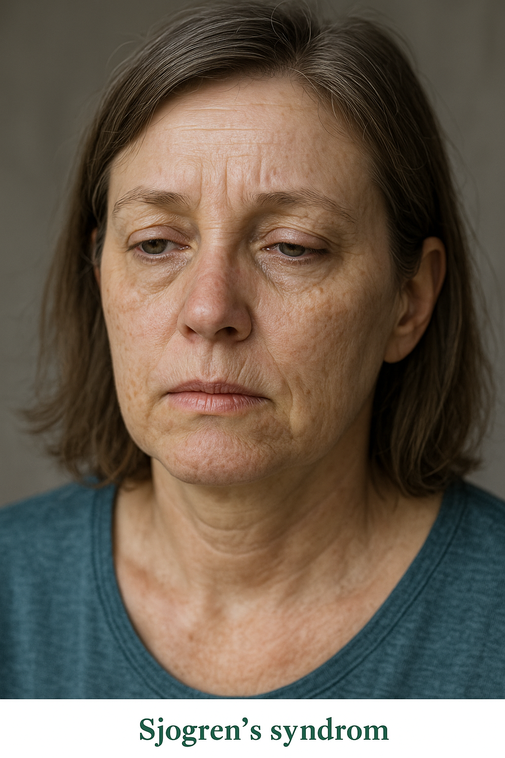
Sjögren’s Syndrome and the Nervous System
What is it?
Sjögren’s syndrome is an autoimmune disease where the body attacks its moisture-producing glands.
It causes dry eyes, dry mouth, and sometimes nerve problems.
Nerve involvement can lead to numbness, tingling, or burning in the hands and feet.
It can also cause facial weakness, dizziness, or memory problems.
Both the central and peripheral nervous systems can be affected.
What causes it?
It happens when the immune system attacks healthy cells by mistake.
The glands that produce tears and saliva are the first to be affected.
In some people, the immune attack also targets nerves.
This may damage the nerve covering (myelin) or the nerve itself.
Genetic and environmental factors play a role in who develops it.
How is it diagnosed?
Diagnosis is based on symptoms like dry eyes and dry mouth, plus blood and nerve tests.
Blood tests look for antibodies called anti-Ro (SSA) and anti-La (SSB).
A small lip biopsy can confirm the disease by showing immune cell clusters.
Nerve tests (EMG, nerve conduction) detect nerve injury or slowing.
MRI may be used if the brain or spinal cord is involved.
What is the treatment?
Treatment aims to control inflammation and relieve symptoms.
Artificial tears and saliva substitutes help dryness.
Medications like steroids, hydroxychloroquine, or immunosuppressants reduce immune activity.
Pain-relieving drugs such as gabapentin or duloxetine help with burning or tingling.
Severe nerve or brain involvement may need stronger medicines like rituximab.
What is the prognosis?
Many people live well with proper treatment and follow-up.
Nerve symptoms may improve slowly and sometimes do not fully recover.
Regular checkups help detect relapses or complications early.
Long-term inflammation increases the risk of lymphoma, so ongoing monitoring is vital.
With good care, most patients maintain an active and independent life.
Small Fiber Neuropathy
What is it?
Small fiber neuropathy (SFN) is a condition where the small nerves that carry pain, temperature, and automatic body signals are damaged.
These nerves include thinly myelinated A\(\delta\) and unmyelinated C fibers.
People often feel burning, tingling, or sharp pain in the feet and hands, especially at night.
It may also affect automatic body functions like sweating, digestion, or blood pressure.
Muscle strength and reflexes usually stay normal, which helps distinguish it from other neuropathies.
What causes it?
Diabetes and impaired glucose tolerance are the most common causes.
Other causes include autoimmune diseases, infections (like HIV or hepatitis C), certain medications, and toxins.
Some people have inherited forms caused by sodium channel gene mutations (e.g., SCN9A, SCN10A).
In up to half of all cases, no clear cause is found (idiopathic SFN).
Chronic inflammation, oxidative stress, and reduced blood supply to nerves contribute to the damage.
How is it diagnosed?
Diagnosis begins with symptoms such as burning pain, tingling, or temperature changes.
Nerve conduction studies are often normal because large nerves are not affected.
A skin biopsy is the gold standard test, showing reduced intraepidermal nerve fiber density.
Other tests include quantitative sensory testing (QST) and autonomic function studies.
Blood tests look for underlying causes like diabetes, autoimmune markers, or vitamin deficiencies.
What is the treatment?
Treating the underlying cause is most important, such as controlling blood sugar in diabetes.
Medications for nerve pain include gabapentin, pregabalin, duloxetine, or amitriptyline.
Topical treatments such as capsaicin or lidocaine patches may help localized pain.
Autonomic symptoms can be managed with fluids, compression stockings, or medicines like midodrine.
Regular follow-up helps monitor symptoms and adjust treatment as needed.
What is the prognosis?
Many patients improve or stabilize with proper treatment.
Pain may persist but can be controlled in most cases.
Identifying and treating the cause early improves outcomes.
Idiopathic cases can be chronic but are usually not life-threatening.
Long-term follow-up ensures symptom control and quality of life.
Spina Bifida
What is it?
Spina bifida is a birth defect where the spine and spinal cord do not close properly during early pregnancy.
It is part of a group of conditions called neural tube defects (NTDs).
In severe cases, the spinal cord and its coverings protrude through an opening in the back.
It can cause weakness, paralysis, or bladder and bowel problems.
The most common and serious type is called myelomeningocele.
What causes it?
It occurs when the neural tube fails to close during the 3rd to 4th week of pregnancy.
Low folic acid levels before or during early pregnancy are a major risk factor.
Other risk factors include certain anti-seizure drugs, diabetes, obesity, and high fever in early pregnancy.
Genetics also play a role, and families with one affected child have a higher risk in future pregnancies.
Environmental and nutritional factors can influence the likelihood of occurrence.
How is it diagnosed?
During pregnancy, blood tests can detect high alpha-fetoprotein (AFP) levels.
Ultrasound scans can show spinal defects or protrusions by 18–20 weeks.
Amniocentesis may show increased acetylcholinesterase activity.
After birth, physical examination and imaging like MRI confirm the diagnosis.
Genetic counseling may be recommended if other abnormalities are seen.
What is the treatment?
Early surgery after birth helps close the defect and prevent infection.
Some cases can be treated with prenatal (in utero) surgery before delivery.
Ongoing care includes physical therapy, urology support, and orthopedic management.
Children may need assistive devices for walking and programs for bladder and bowel care.
Folic acid supplements before and during pregnancy can prevent most cases.
What is the prognosis?
Many children live fulfilling lives with proper treatment and rehabilitation.
The level of the spinal defect determines how much movement or sensation is affected.
Early surgery and multidisciplinary care improve long-term outcomes.
Some may develop hydrocephalus requiring a shunt to drain excess brain fluid.
Lifelong follow-up ensures mobility, continence, and independence.
Spinal Cord Injury
What is it?
A spinal cord injury (SCI) happens when the spinal cord is damaged by trauma or disease.
It can cause partial or complete loss of movement and sensation below the level of injury.
Common causes include vehicle accidents, falls, sports injuries, or violence.
The injury may also affect bladder, bowel, and blood pressure control.
Depending on where the injury occurs, it may cause paraplegia (legs only) or quadriplegia (arms and legs).
What causes it?
Most SCIs are caused by trauma that fractures or dislocates the spine.
Other causes include tumors, infections, bleeding, or inflammation around the spinal cord.
The initial mechanical injury is followed by secondary damage from swelling, poor blood flow, and cell death.
This cascade worsens nerve injury and limits recovery if not treated promptly.
Early stabilization of the spine helps prevent further damage.
How is it diagnosed?
Doctors assess muscle strength, sensation, and reflexes to find the level of injury.
MRI scans show spinal cord compression, swelling, or bleeding.
CT scans help detect bone fractures or dislocations.
The ASIA Impairment Scale is used to grade the severity of injury.
Other tests check bladder, bowel, and autonomic function.
What is the treatment?
Immediate care includes stabilizing the spine and supporting breathing and blood pressure.
Surgery may be needed to decompress the spinal cord and fix fractures.
Rehabilitation starts early to preserve muscle strength and prevent complications.
Medications and physical therapy help manage spasticity and pain.
Long-term care focuses on mobility aids, bladder management, and skin protection.
What is the prognosis?
Recovery depends on the level and completeness of injury.
Partial injuries often have better outcomes than complete injuries.
Rehabilitation and assistive technology can restore independence in many cases.
Complications like infections and pressure ulcers can be prevented with care.
Emotional and social support play a key role in long-term adjustment and quality of life.
Spinal Muscular Atrophy (SMA)
What is it?
Spinal muscular atrophy (SMA) is a genetic condition that causes muscles to weaken and shrink (atrophy) over time.
It occurs because the nerve cells in the spinal cord that control movement (motor neurons) are lost.
The brain remains unaffected, so intelligence and thinking ability are normal.
There are several types depending on the age of onset and severity, from severe infantile forms to mild adult-onset disease.
Without treatment, severe forms can affect breathing and swallowing early in life.
What causes it?
SMA is caused by a defect in the SMN1 gene on chromosome 5.
This gene makes a protein essential for motor neuron survival.
When both SMN1 genes are faulty, motor neurons die and muscles lose function.
A backup gene, SMN2, makes some protein but not enough; having more SMN2 copies usually means milder symptoms.
It is inherited in an autosomal recessive pattern—both parents must carry one defective gene.
How is it diagnosed?
A genetic blood test confirms the diagnosis by detecting deletion or mutation of the SMN1 gene.
Doctors may also assess muscle tone, reflexes, and movement abilities.
Nerve and muscle tests (EMG or nerve conduction studies) can show reduced motor neuron activity.
Muscle biopsy is rarely needed today since genetic testing is highly accurate.
Many countries now include SMA in newborn screening programs, allowing early diagnosis before symptoms appear.
What is the treatment?
New treatments can dramatically improve survival and muscle function if started early.
Nusinersen (Spinraza) helps the body make more SMN protein from the SMN2 gene.
Onasemnogene abeparvovec (Zolgensma) replaces the missing SMN1 gene through gene therapy.
Risdiplam (Evrysdi) is an oral medicine that also increases SMN protein production.
Supportive care includes physiotherapy, breathing support, nutrition management, and orthopedic care for posture and scoliosis.
What is the prognosis?
With modern therapies, many children with SMA live longer and gain motor skills previously thought impossible.
Early diagnosis and treatment are key for the best outcomes.
Ongoing physiotherapy helps maintain mobility and joint flexibility.
Severe forms may still need respiratory and feeding support.
With comprehensive care, most patients lead longer, more independent lives than before.
Stiff Person Syndrome (SPS)
What is it?
Stiff Person Syndrome (SPS) is a rare neurological disorder that causes muscle stiffness and painful spasms.
It happens because the body’s immune system attacks parts of the brain and spinal cord that control muscle movement.
The stiffness mainly affects the back and leg muscles, leading to a rigid posture and difficulty walking.
Spasms can be triggered by sudden noise, touch, or emotional stress.
Without treatment, stiffness can worsen and limit daily activities.
What causes it?
SPS is an autoimmune disorder in which the body attacks its own nerve cells.
Most people with SPS have antibodies against an enzyme called GAD65, which helps make GABA—a chemical that calms nerve activity.
Without enough GABA, muscles stay overactive and stiff.
Some cases are linked to cancers (paraneoplastic SPS), especially breast or lung cancer.
It is more common in women and often occurs alongside other autoimmune diseases like diabetes or thyroiditis.
How is it diagnosed?
Diagnosis is based on symptoms, examination, and special tests.
Electromyography (EMG) shows continuous muscle activity even at rest.
A blood test can detect anti-GAD65 or other antibodies related to the disorder.
MRI scans are usually normal but help rule out other causes.
Sometimes doctors look for hidden cancers if a paraneoplastic form is suspected.
What is the treatment?
Medicines that boost GABA levels, like diazepam or baclofen, help relax muscles and reduce stiffness.
Intravenous immunoglobulin (IVIG) or plasmapheresis can reduce harmful antibodies.
Immunosuppressants such as steroids or rituximab may help in more severe or persistent cases.
Physical therapy is vital to improve flexibility, prevent contractures, and support walking.
Treating an underlying cancer (if present) can also improve symptoms.
What is the prognosis?
SPS often improves with treatment, especially when started early.
Many people can manage symptoms and maintain independence with medication and therapy.
Some patients experience relapses, requiring long-term immunotherapy.
Severe cases may affect breathing or cause disability without care.
With proper treatment and support, most patients can live active, fulfilling lives.
Stroke - Cardioembolic
What is it?
A cardioembolic stroke occurs when a blood clot forms in the heart and travels to the brain, blocking blood flow to part of the brain.
This type of stroke usually happens suddenly and often causes more severe symptoms than other stroke types.
The most common cause is atrial fibrillation, a heart rhythm problem that allows blood clots to form inside the heart.
The clot then moves through the bloodstream and lodges in a brain artery, cutting off oxygen to brain tissue.
Without rapid treatment, this can lead to permanent brain injury or disability.
What causes it?
Cardioembolic stroke is caused by a clot that starts in the heart and travels to the brain.
Common causes include:
Atrial fibrillation (AF): irregular heartbeat causing blood to pool and clot in the atrium.
Heart attack or heart failure: damaged heart muscle can lead to clots in areas where the heart does not pump effectively.
Heart valve problems or artificial valves: increase risk of clot formation.
Infective endocarditis: infection on heart valves can produce fragments that break off and travel to the brain.
Patent foramen ovale (PFO): a small hole in the heart that allows clots from veins to pass into arteries.
How is it diagnosed?
Brain imaging with CT or MRI confirms a stroke and shows the affected brain area.
Echocardiography (ultrasound of the heart) identifies blood clots, valve problems, or heart wall abnormalities.
ECG or continuous cardiac monitoring detects atrial fibrillation or other irregular rhythms.
Blood tests check for clotting problems, infection, or other contributing factors.
Doctors may also use vascular imaging (CT or MR angiography) to ensure that arteries are open and rule out other causes.
What is the treatment?
Emergency treatment focuses on restoring blood flow and preventing further damage.
Clot-busting medication (thrombolysis) can dissolve the clot if given within 4.5 hours of symptom onset.
Mechanical thrombectomy can remove the clot directly from large arteries within 6–24 hours in selected cases.
Once bleeding risk is excluded, anticoagulants (such as apixaban, rivaroxaban, dabigatran, or warfarin) are prescribed to prevent new clots.
Blood pressure, cholesterol, and diabetes must be controlled to reduce recurrence risk.
Treatment of underlying heart conditions (like rhythm control for AF or valve repair) is also essential.
What is the prognosis?
Recovery depends on the size and location of the stroke and how quickly treatment was started.
With proper anticoagulation, the risk of a second stroke can be reduced by up to 70%.
Early rehabilitation helps regain strength, speech, and mobility.
Some patients may have lasting weakness or difficulty speaking but can improve with therapy.
Lifelong follow-up with a neurologist and cardiologist is recommended to prevent recurrence and manage heart rhythm or valve issues.
Stroke - Small Vessel (Lacunar Stroke)
What is it?
A small vessel or lacunar stroke occurs when one of the tiny arteries deep inside the brain becomes blocked.
These small strokes usually affect structures like the internal capsule, thalamus, or brainstem.
They are often mild but can still cause weakness, speech changes, or balance problems.
Because they affect small areas, symptoms may be limited but can add up over time if multiple strokes occur.
It is one of the most common types of ischemic stroke, especially in people with long-standing high blood pressure or diabetes.
What causes it?
Small vessel strokes are caused by damage to the tiny penetrating arteries in the brain.
The most common reason is long-term high blood pressure, which weakens and thickens the walls of these vessels.
Diabetes also contributes by damaging the blood vessel lining.
Over time, these changes cause narrowing (lipohyalinosis) and blockage of blood flow.
Less often, small cholesterol plaques or microemboli from larger arteries can block these small vessels.
How is it diagnosed?
Diagnosis is based on symptoms, imaging, and exclusion of other stroke types.
MRI, especially diffusion-weighted imaging (DWI), is the best test—it shows small, deep infarcts usually less than 15 mm wide.
CT scans may appear normal early but can show small areas of damage later.
Vascular imaging (CT or MR angiography) rules out blockages in larger arteries.
Cardiac studies and blood tests help ensure there is no heart-related or clotting cause.
What is the treatment?
Immediate care follows standard ischemic stroke management, including evaluation for clot-busting therapy if eligible.
Antiplatelet drugs (such as aspirin or clopidogrel) are used to prevent future strokes.
Blood pressure control is the most important step in preventing recurrence.
Good management of diabetes, cholesterol, and lifestyle factors—like quitting smoking and regular exercise—helps protect brain blood vessels.
Rehabilitation, including physiotherapy and speech therapy, promotes recovery from weakness or speech problems.
What is the prognosis?
Most people recover well, especially if only one small vessel is affected.
However, repeated small strokes can cause problems with walking, balance, or memory over time.
Strict control of blood pressure and diabetes greatly reduces the risk of future events.
Some patients may develop “vascular dementia” if many small strokes occur over years.
Early diagnosis, lifestyle changes, and medication adherence lead to the best long-term outcome.
Syringomyelia
What is it?
Syringomyelia is a condition where a fluid-filled cavity (called a syrinx) forms inside the spinal cord.
This cavity can slowly get bigger and damage the nerves in the spinal cord.
It often causes problems with feeling, especially pain and temperature, in the arms and hands.
Muscle weakness and wasting, especially in the hands, are common.
Symptoms usually develop slowly over months or years.
What causes it?
The most common cause is a problem at the base of the brain called Chiari malformation, where brain tissue pushes into the spinal canal.
Past injuries to the spine, such as trauma or surgery, can also lead to syringomyelia.
Tumors or growths in the spinal cord may block fluid flow and cause a syrinx to form.
Infections or inflammation around the spinal cord can sometimes be a cause.
In some cases, no clear cause is found (idiopathic syringomyelia).
How is it diagnosed?
Doctors start with a physical exam and ask about symptoms like numbness, weakness, or pain.
MRI (magnetic resonance imaging) of the spine is the main test to see the syrinx and any related problems.
Sometimes, special MRI scans are done to see how spinal fluid flows.
Other tests may be used to look for causes, such as tumors or Chiari malformation.
Nerve and muscle tests (EMG) can help check how the nerves are working.
What is the treatment?
Treatment depends on the cause and how severe the symptoms are.
Surgery may be needed to correct the underlying problem, such as Chiari malformation or to drain the syrinx.
Some people with mild or stable symptoms are monitored with regular MRI scans and checkups.
Physical therapy and pain management can help with muscle weakness and discomfort.
Treating the underlying cause, when possible, can stop the syrinx from getting worse.
What is the prognosis?
Many people see improvement or stabilization of symptoms after treatment, especially if treated early.
Some nerve damage may be permanent if the syrinx has been present for a long time.
Regular follow-up is important to monitor for changes or new symptoms.
Early diagnosis and treatment give the best chance for a good outcome.
With proper care, many patients can lead active and fulfilling lives.
Tardive Dyskinesia
What is it?
Tardive dyskinesia is a condition that causes involuntary, repetitive movements, often of the face, mouth, or limbs.
It usually develops after taking certain medications for a long time, especially those used to treat mental health conditions.
The movements can include lip smacking, tongue movements, grimacing, or rapid blinking.
These movements are not under the person’s control and can be mild or severe.
Tardive dyskinesia can affect daily activities and may be distressing to those who have it.
What causes it?
The main cause is long-term use of medications that block dopamine, a brain chemical involved in movement.
Medicines most commonly linked to tardive dyskinesia include antipsychotics and some drugs for nausea.
The risk increases the longer someone takes these medications and at higher doses.
Older adults and women may be at higher risk.
Sometimes, symptoms can continue even after stopping the medication.
How is it diagnosed?
Diagnosis is based on noticing the typical movements and a history of taking certain medications.
There are no special blood tests or scans needed for diagnosis.
Doctors may use a movement rating scale to track symptoms over time.
Other conditions with similar symptoms may need to be ruled out.
Early recognition is important to help manage the condition.
What is the treatment?
The first step is to review and possibly adjust the medications causing the symptoms.
Sometimes, switching to a different medicine can help reduce the movements.
There are specific medications approved to treat tardive dyskinesia and help control the movements.
Other treatments, like certain muscle relaxants, may be tried if needed.
Regular doctor visits are important to monitor and manage symptoms.
What is the prognosis?
Some people improve after changing or stopping the medication, but symptoms can last a long time.
In some cases, the movements may become less noticeable over time.
For others, the symptoms can be persistent and affect quality of life.
Early detection and treatment can improve outcomes.
Support from healthcare providers, family, and friends can help patients cope with the condition.
Tarsal Tunnel Syndrome
What is it?
Tarsal tunnel syndrome is a condition where a nerve in the ankle gets squeezed or compressed.
This nerve, called the tibial nerve, passes through a narrow space near the inside of the ankle.
Compression can cause pain, tingling, or numbness in the foot and toes.
Symptoms often get worse with standing or walking for long periods.
It is similar to carpal tunnel syndrome but affects the ankle and foot instead of the wrist and hand.
What causes it?
Tarsal tunnel syndrome is usually caused by anything that puts pressure on the tibial nerve.
Causes can include flat feet, swelling, injuries, or growths like cysts near the ankle.
Medical conditions like diabetes or arthritis can increase the risk.
Repetitive activities or standing for long times may contribute.
Sometimes, the cause is not found.
How is it diagnosed?
Doctors ask about symptoms and examine the foot and ankle.
Tapping over the nerve (Tinel’s sign) may cause tingling or pain.
Special nerve tests can show if the nerve is being compressed.
Imaging like ultrasound or MRI may be used to look for causes of compression.
Other foot problems may need to be ruled out before making the diagnosis.
What is the treatment?
Treatment usually starts with rest, changing activities, and wearing supportive shoes or inserts.
Medicines like anti-inflammatories can help reduce pain and swelling.
Physical therapy may help improve symptoms.
In some cases, injections or surgery may be needed if symptoms do not improve.
Treating any underlying cause, such as removing a cyst or controlling diabetes, is important.
What is the prognosis?
Many people improve with simple treatments and activity changes.
If treated early, long-term nerve damage can often be prevented.
Some people may need surgery if symptoms are severe or do not get better.
Recovery after surgery is usually good, but it may take time.
Ongoing support and foot care can help prevent symptoms from returning.
Temporal Arteritis (Giant Cell Arteritis)
What is it?
Temporal arteritis, also called giant cell arteritis, is an inflammation of the blood vessels, mainly those in the head.
It most often affects adults over 50 years old.
The condition can cause headaches, scalp tenderness, and jaw pain.
It may lead to vision problems if not treated quickly.
It is considered a medical emergency because of the risk of permanent vision loss.
What causes it?
The exact cause is not known, but it is thought to involve the immune system attacking blood vessel walls.
Age is a major risk factor, as it mainly affects older adults.
Genetics and environmental factors may play a role.
It may sometimes occur with another condition called polymyalgia rheumatica.
There is no evidence that lifestyle or diet alone causes the disease.
How is it diagnosed?
Doctors diagnose it based on symptoms like headache, jaw pain, and vision changes.
Blood tests can show signs of inflammation, such as a high ESR or CRP.
A biopsy of the temporal artery may be done to confirm the diagnosis.
Imaging tests like ultrasound or MRI can help detect inflamed arteries.
Quick diagnosis is important to prevent vision loss.
What is the treatment?
Treatment usually starts right away with high-dose steroids (like prednisone) to reduce inflammation.
Sometimes, other medicines are added to help lower the steroid dose and control the disease.
Low-dose aspirin may be recommended to reduce the risk of blood clots.
Regular follow-up is needed to monitor symptoms and side effects of treatment.
Treatment may last for a year or longer, with gradual reduction of medication.
What is the prognosis?
Most people respond well to treatment if started early.
Vision loss can be permanent if treatment is delayed.
Some people may have relapses and need longer treatment.
Side effects from steroids are possible, so regular check-ups are important.
With proper care, most people can return to normal activities.
Temporomandibular Joint (TMJ) Syndrome
What is it?
TMJ syndrome is a disorder affecting the jaw joint and the muscles that control jaw movement.
It often causes pain or discomfort in the jaw, face, or around the ear.
People may notice clicking, popping, or grating sounds when opening or closing the mouth.
Jaw stiffness or trouble opening the mouth wide is common.
It can affect one or both sides of the jaw.
What causes it?
TMJ syndrome can be caused by jaw injury or arthritis in the joint.
Teeth grinding or clenching (especially during sleep) can strain the jaw joint.
Stress, which leads to jaw muscle tension, may contribute.
Poor bite alignment or missing teeth can increase risk.
Sometimes, the exact cause is not known.
How is it diagnosed?
Doctors ask about symptoms and examine the jaw for tenderness, movement, and sounds.
They may check how wide you can open your mouth and feel for clicking or popping.
Dental x-rays or other imaging (like MRI) may be used if needed.
Diagnosis is often made based on symptoms and examination alone.
Other causes of facial pain are ruled out before making the diagnosis.
What is the treatment?
Most cases improve with simple measures like rest, a soft diet, and avoiding wide jaw movements.
Applying warm compresses or gentle jaw exercises may help relieve pain.
Medicines such as pain relievers or muscle relaxants can be used if needed.
A dentist may suggest a mouth guard or splint, especially if you grind your teeth.
Surgery is rarely needed and is only considered if other treatments do not help.
What is the prognosis?
Most people get better with simple treatments and time.
Symptoms may come and go, but are usually not serious.
Managing stress and avoiding jaw overuse can help prevent flare-ups.
Long-term problems are uncommon if treated early.
Ongoing dental care and follow-up can help keep symptoms under control.
Thoracic Outlet Syndrome
What is it?
Thoracic outlet syndrome is a condition where nerves or blood vessels are squeezed as they pass from the neck to the arm.
It can cause pain, numbness, tingling, or weakness in the arm or hand.
Some people may notice swelling or color changes in the arm.
Symptoms often get worse with certain arm positions or activities.
There are three main types: nerve, vein, and artery compression, with nerve type being the most common.
What causes it?
It can be caused by extra ribs, tight muscles, or abnormal bands of tissue in the neck or shoulder.
Repetitive overhead movements or injuries can increase the risk.
Poor posture or carrying heavy loads may contribute.
Some people are born with anatomy that makes them more likely to develop this condition.
Sometimes, the exact cause is not found.
How is it diagnosed?
Doctors ask about symptoms and examine the neck, shoulder, and arm.
Certain arm positions or movements during the exam may reproduce symptoms.
Imaging tests like ultrasound, MRI, or nerve studies may be done to look for compression.
Blood flow tests can help if swelling or color changes are present.
Other conditions with similar symptoms are ruled out before diagnosis.
What is the treatment?
Treatment often starts with physical therapy to improve posture and strengthen shoulder muscles.
Avoiding activities that trigger symptoms is important.
Pain relievers or anti-inflammatory medicines may help manage discomfort.
In some cases, surgery is needed to relieve pressure on nerves or blood vessels.
Ongoing care and exercises can help prevent symptoms from returning.
What is the prognosis?
Many people improve with therapy and activity changes.
Early treatment can prevent long-term nerve or blood vessel damage.
Some may need surgery, especially if symptoms are severe or do not improve.
After treatment, most people can return to normal activities.
Regular follow-up helps ensure symptoms stay under control.
Tics
What is it?
Tics are sudden, quick movements or sounds that a person does over and over again.
These movements or sounds are hard to control and may happen many times a day.
Tics can involve blinking, facial grimacing, shoulder shrugging, or making noises like throat clearing.
They often start in childhood, most commonly between ages 4 and 6.
Tics may come and go, and their severity can change over time.
What causes it?
The exact cause of tics is not fully understood, but both genes and environment play a role.
Tics often run in families, suggesting a genetic link.
Stress, excitement, or fatigue can make tics more noticeable or frequent.
Some infections or changes in brain chemicals may also contribute.
Most tics are not caused by anything a child or parent did.
How is it diagnosed?
Doctors diagnose tics by asking about symptoms and watching the movements or sounds.
No special blood tests or scans are usually needed.
The doctor will check how long the tics have been present and how they affect daily life.
It is important to rule out other conditions that may look similar to tics.
Early diagnosis can help families understand and manage tics better.
What is the treatment?
Many children with mild tics do not need treatment, just reassurance and support.
Behavioral therapy, such as habit reversal training, can help children control bothersome tics.
Medicines may be used if tics are severe or interfere with daily activities.
Treating other conditions like ADHD or anxiety can also help improve tics.
Regular follow-up with the doctor helps track symptoms and adjust care as needed.
What is the prognosis?
Most children with tics improve as they get older, and many outgrow them by adolescence.
Tics may come and go, and sometimes return during times of stress.
Some people continue to have tics into adulthood, but they are often milder.
Support from family, teachers, and friends helps children cope with tics.
Early understanding and treatment can lead to better outcomes and less worry.
Tinnitus
What is it?
Tinnitus is the hearing of sounds, such as ringing or buzzing, when there is no external noise present.
The sound may be heard in one or both ears, or inside the head.
It can be constant or come and go, and may be soft or loud.
Many people notice tinnitus more in quiet settings or at night.
Tinnitus is a common problem and can affect people of all ages.
What causes it?
Tinnitus can happen when there is damage to the tiny hair cells in the inner ear, often from loud noise or aging.
Ear infections, earwax buildup, or certain medications can also cause tinnitus.
Conditions like hearing loss, head injuries, or problems with blood vessels may play a role.
Sometimes, stress or anxiety can make tinnitus more noticeable.
In many cases, the exact cause is not found.
How is it diagnosed?
The doctor will ask about your symptoms and medical history.
An ear exam will be done to check for ear problems.
Hearing tests are often used to see if there is any hearing loss.
Sometimes, imaging tests may be needed if the tinnitus is only in one ear or is pulsating.
The doctor may ask about medicines you take or recent illnesses to help find the cause.
What is the treatment?
Treating any underlying ear problems, such as removing wax or treating infections, can help.
Hearing aids may help if you have hearing loss along with tinnitus.
Sound therapy or using background noise can make tinnitus less noticeable.
Counseling or cognitive behavioral therapy can help you cope with the stress or anxiety tinnitus may cause.
Medicines are rarely used, but may help if you have trouble sleeping or feel very anxious.
What is the prognosis?
For many people, tinnitus does not get worse and may improve over time.
Learning ways to manage tinnitus can help reduce its impact on daily life.
Most people can continue normal activities with little or no trouble.
Tinnitus can be bothersome, but serious complications are rare.
Support from healthcare providers, family, and friends can make coping with tinnitus easier.
Tolosa–Hunt Syndrome
What is it?
Tolosa–Hunt Syndrome is a rare cause of sudden, painful eye movement due to inflammation around the eye nerves.
It often causes severe pain behind one eye, followed by weakness or paralysis of the eye muscles.
The pain may spread to nearby areas like the forehead or temple.
The condition affects the area called the cavernous sinus or orbital apex, where many eye nerves pass.
It is not an infection or cancer, but an inflammation that can mimic more serious conditions.
What causes it?
The exact cause is unknown, but it is believed to result from an autoimmune inflammation.
The immune system attacks tissues around the nerves controlling eye movement.
It is not linked to trauma or infection.
Rarely, it may follow or be confused with other conditions like sarcoidosis or vasculitis.
Stress or immune reactions might trigger the inflammation in some individuals.
How is it diagnosed?
Diagnosis is based on symptoms of eye pain and double vision, along with imaging studies.
MRI of the brain and orbit often shows inflammation near the eye nerves.
Blood tests help rule out infections or autoimmune diseases.
Other causes like tumors or aneurysms must be excluded first.
Rapid response to steroids strongly supports the diagnosis.
What is the treatment?
Corticosteroids are the main treatment and usually bring quick relief from pain.
The dose is gradually reduced over weeks to months to prevent relapse.
If symptoms return, another short course of steroids may be needed.
In rare cases, other immune-suppressing medicines may be added.
Pain control and follow-up MRI are important parts of management.
What is the prognosis?
Most people recover completely with proper treatment.
Eye movements and pain usually improve within days to weeks.
Some may experience occasional relapses that respond to steroids.
Permanent eye nerve weakness is uncommon.
Long-term outlook is generally excellent with early recognition and care.
Tourette Syndrome
What is it?
Tourette Syndrome is a condition that causes sudden, repeated movements or sounds called tics.
These can include blinking, shrugging, sniffing, or throat clearing.
Tics often start in childhood and may change over time.
They can get worse with stress and lessen during calm or focused activities.
The condition is not dangerous, but it can affect school or social life.
What causes it?
It results from changes in how the brain controls movement and habits.
The circuits involving dopamine and other brain chemicals are overactive.
Genetics play a major role, and it often runs in families.
Stress, fatigue, or infections may temporarily worsen symptoms.
It is not caused by parenting style or emotional weakness.
How is it diagnosed?
Diagnosis is based on observing tics and their pattern over time.
No specific blood test or scan confirms it.
Doctors look for both motor (movement) and vocal (sound) tics lasting more than a year.
Other conditions like seizures or stereotypies are ruled out.
A neurologist or movement disorder specialist usually makes the diagnosis.
What is the treatment?
Many children need only reassurance and education.
Behavioral therapy such as CBIT can help manage tics.
Medicines like risperidone or clonidine may be used for severe tics.
Treating associated conditions such as ADHD or anxiety is important.
Support from teachers and family improves coping and confidence.
What is the prognosis?
Tics often peak in early adolescence and improve with age.
Many people have fewer or no tics as adults.
Emotional support helps children adapt and thrive.
Recurrences can happen during stress but are usually mild.
Long-term outcomes are excellent for most individuals.
Transient Ischemic Attack (TIA)
What is it?
A Transient Ischemic Attack, or TIA, is often called a “mini-stroke.”
It happens when blood flow to part of the brain is briefly blocked.
Symptoms are similar to a stroke but last only minutes to hours.
Common symptoms include weakness, speech difficulty, or vision loss.
It is a serious warning sign for a future stroke.
What causes it?
TIA occurs due to a temporary blockage in a brain artery, often from a small blood clot.
Risk factors include high blood pressure, diabetes, smoking, and high cholesterol.
Irregular heart rhythms like atrial fibrillation can send clots to the brain.
Narrowing of neck arteries (carotid stenosis) also increases risk.
Poor lifestyle habits and older age make TIAs more likely.
How is it diagnosed?
TIA is diagnosed by evaluating symptoms and doing brain scans.
MRI or CT helps rule out a full stroke or bleeding.
Carotid ultrasound and heart tests identify possible sources of clots.
Blood work checks for cholesterol, sugar, and clotting problems.
Doctors may observe patients briefly, even if symptoms have resolved.
What is the treatment?
Treatment focuses on preventing a future stroke.
Blood thinners like aspirin or clopidogrel are commonly prescribed.
Blood pressure, sugar, and cholesterol control are essential.
Lifestyle changes such as quitting smoking and regular exercise are vital.
Surgery or stenting may be done if neck arteries are severely narrowed.
What is the prognosis?
A TIA is reversible, but it is a major warning sign for stroke.
The highest risk of stroke is within days after a TIA.
Prompt evaluation and treatment greatly reduce this risk.
With proper care, most people recover fully and prevent future events.
Regular follow-up and healthy habits protect long-term brain health.
Transverse Myelitis
What is it?
Transverse myelitis is an inflammation of the spinal cord that causes sudden weakness, numbness, and bladder problems.
It affects both sides of the body below a certain level, depending on where the spinal cord is inflamed.
The condition can come on quickly over hours or days.
It is not an infection but an immune reaction that damages the nerve fibers in the spine.
Early treatment can help prevent long-term disability.
What causes it?
It can occur after viral or bacterial infections that trigger an immune response.
Some cases are linked to autoimmune diseases such as multiple sclerosis or neuromyelitis optica.
In many people, the exact cause is not found.
Rarely, it can happen after vaccination or other immune stimulation.
The inflammation damages the protective myelin around spinal nerves.
How is it diagnosed?
MRI scans show inflammation or swelling within the spinal cord.
A spinal tap (lumbar puncture) checks for infection and immune activity in the fluid.
Blood tests may look for antibodies linked to specific disorders like NMOSD or MOG disease.
Other conditions such as stroke, compression, or tumor are ruled out with imaging.
Doctors assess strength, sensation, and reflexes to localize the spinal level affected.
What is the treatment?
High-dose steroids are given early to reduce inflammation and swelling.
If symptoms do not improve, plasma exchange may be used to remove harmful antibodies.
Some patients may need longer-term immune therapy to prevent relapses.
Physical and occupational therapy help restore movement and independence.
Pain control, bladder training, and support care are key parts of recovery.
What is the prognosis?
Many patients recover well, especially with early treatment.
Some may be left with mild weakness or numbness.
Severe cases can lead to lasting disability or need for mobility aids.
Follow-up is important to monitor for recurrence or conversion to multiple sclerosis.
With rehabilitation and support, most people regain significant function.
Trigeminal Neuralgia
What is it?
Trigeminal neuralgia is a nerve condition that causes sudden, sharp, electric shock-like pain in the face.
It affects one side of the face, usually the cheek or jaw.
Pain can be triggered by light touch, talking, or brushing teeth.
Episodes are brief but can happen many times a day.
It is sometimes called the “suicide disease” because of its intensity, though it can be treated effectively.
What causes it?
The main cause is pressure on the trigeminal nerve by a nearby blood vessel.
This pressure damages the nerve covering (myelin), causing it to misfire.
In some cases, it is related to multiple sclerosis or a tumor pressing on the nerve.
Injury or infection in the face may also play a role.
Often, no clear cause is found, especially in older adults.
How is it diagnosed?
Diagnosis is based on the description of pain—brief, sharp, one-sided, and trigger-sensitive.
The doctor will check the face and nerve function.
MRI scans are done to rule out tumors or multiple sclerosis.
Tests may help confirm nerve compression or exclude dental or sinus problems.
Response to medications like carbamazepine helps confirm the diagnosis.
What is the treatment?
First-line treatment is medication that calms overactive nerves, such as carbamazepine or oxcarbazepine.
If these fail, other drugs like gabapentin or baclofen can be tried.
Surgery may relieve pressure on the nerve (microvascular decompression).
For those who cannot have surgery, procedures like radiosurgery or nerve blocks may help.
Lifestyle changes and stress management can reduce pain triggers.
What is the prognosis?
Many patients respond very well to medication.
Surgical options provide long-lasting relief for most who need them.
Recurrence may happen but can often be controlled again with treatment.
It is not life-threatening, but chronic pain can affect mood and quality of life.
With proper care, most people live normally and pain-free.
Trochlear Nerve (Cranial Nerve IV) Palsy
What is it?
Trochlear nerve palsy is a weakness of one eye muscle that causes double vision.
It affects the superior oblique muscle, which helps the eye move downward and inward.
People often notice vertical double vision, especially when looking down.
To compensate, they may tilt their head to one side.
It can be temporary or permanent, depending on the cause.
What causes it?
The most common cause is small blood vessel damage from diabetes or high blood pressure.
Head injury can stretch or damage the delicate trochlear nerve.
It may also occur with brain tumors, inflammation, or stroke.
Some people are born with it and only notice symptoms later in life.
Rarely, it can follow surgery or infection near the brainstem.
How is it diagnosed?
Eye movement testing helps identify the affected muscle.
The Bielschowsky head tilt test confirms the diagnosis by worsening the double vision on head tilt.
MRI or CT scans may be ordered to rule out trauma or structural causes.
Blood sugar and blood pressure are checked for small-vessel disease.
The doctor examines for signs of other nerve involvement.
What is the treatment?
Many cases recover on their own within a few months.
Prism glasses can reduce double vision during recovery.
Treating underlying problems like diabetes helps prevent recurrence.
Persistent cases may need eye muscle surgery for alignment.
Eye exercises and regular follow-up support visual recovery.
What is the prognosis?
Most people recover fully, especially if the cause is small-vessel disease or mild trauma.
Recovery usually happens within 3–6 months.
Surgery or prism glasses can correct lasting double vision.
Recurrence is uncommon once the cause is treated.
Early diagnosis helps avoid long-term visual strain and discomfort.
Ulnar Neuropathy at Elbow
What is it?
Ulnar neuropathy at the elbow is a condition where the ulnar nerve gets compressed or irritated near the elbow.
This nerve controls feeling in the ring and little fingers and some hand movements.
Compression causes numbness, tingling, and sometimes weakness in the hand.
It is sometimes called cubital tunnel syndrome.
Symptoms often get worse when the elbow is bent for a long time.
What causes it?
Pressure on the ulnar nerve from leaning on the elbow or bending it for long periods.
Repetitive movements or activities that involve frequent elbow bending.
Injuries or swelling around the elbow that squeeze the nerve.
Certain medical conditions like diabetes or obesity can increase risk.
Anatomical differences or changes in the elbow area can also cause nerve irritation.
How is it diagnosed?
A doctor will ask about symptoms and examine the arm and hand.
Tests like nerve conduction studies measure how well the nerve sends signals.
Electromyography (EMG) checks the muscles controlled by the nerve.
Sometimes ultrasound or MRI is used to look at the nerve and surrounding tissues.
Diagnosis is based on symptoms, physical exam, and test results.
What is the treatment?
Avoid activities that put pressure on the elbow or require prolonged bending.
Use a splint or brace to keep the elbow straight, especially at night.
Take anti-inflammatory medications to reduce pain and swelling.
Physical therapy can help improve nerve and muscle function.
Surgery may be needed if symptoms do not improve or if there is significant weakness.
What is the prognosis?
Many people improve with early treatment and lifestyle changes.
Mild cases often get better without surgery.
Surgery usually helps relieve symptoms and prevent further nerve damage.
Recovery time varies but can take several weeks to months.
If untreated, the nerve damage can become permanent, causing lasting weakness or numbness.
Ulnar Neuropathy at the Wrist
What is it?
Ulnar neuropathy at the wrist is when the ulnar nerve is compressed near the wrist.
This nerve controls feeling in the ring and little fingers and some hand movements.
Compression causes numbness, tingling, and weakness in the hand.
It is also called Guyon’s canal syndrome.
Symptoms usually affect the hand but not the forearm.
What causes it?
Repeated pressure on the wrist, such as from cycling or using hand tools.
Injuries or swelling near the wrist that press on the nerve.
Growths like cysts or tumors in the wrist area.
Certain wrist positions held for long periods.
Anatomical differences that make the nerve more likely to be compressed.
How is it diagnosed?
Doctors ask about symptoms and examine the hand and wrist.
Nerve conduction studies check how well the nerve sends signals at the wrist.
Electromyography (EMG) looks at the muscles controlled by the nerve.
Imaging tests like ultrasound or MRI can show any cysts or abnormalities.
Diagnosis is based on symptoms, physical exam, and test results.
What is the treatment?
Avoid activities that put pressure on the wrist or cause symptoms.
Use wrist splints to keep the wrist in a neutral position.
Take anti-inflammatory medicines to reduce pain and swelling.
Ergonomic changes, like padded gloves or changing hand positions, can help.
Surgery may be needed to remove cysts or relieve pressure if symptoms persist.
What is the prognosis?
Many people improve with rest and conservative treatment.
Early treatment can prevent permanent nerve damage.
Surgery often helps if conservative measures don’t work.
Recovery can take weeks to months depending on severity.
Without treatment, symptoms may worsen and lead to lasting weakness or numbness.
Vasculitis of CNS
What is it?
Vasculitis of the CNS is inflammation of blood vessels in the brain and spinal cord.
It can cause problems with how the brain works, such as headaches or weakness.
This condition is rare and can affect people of any age.
It is sometimes called "primary CNS vasculitis" if it only affects the brain.
Symptoms can be different for each person and may come on slowly or suddenly.
What causes it?
The exact cause is often unknown.
Sometimes, the immune system attacks the blood vessels by mistake.
Infections or other diseases can trigger this inflammation.
Some medicines may rarely cause it.
It is not usually inherited or passed down in families.
How is it diagnosed?
Doctors ask about symptoms and do a physical and neurological exam.
Brain scans like MRI are used to look for signs of inflammation or stroke.
Spinal fluid may be tested with a lumbar puncture.
Sometimes, a small piece of brain tissue (biopsy) is needed for confirmation.
Blood tests are done to rule out other causes.
What is the treatment?
Treatment usually starts with strong medicines to reduce inflammation, like steroids.
Other medications may be added to help control the immune system.
Treatment is tailored to each person’s symptoms and severity.
Regular check-ups and follow-up scans help monitor progress.
Early treatment is important to prevent long-term problems.
What is the prognosis?
Many people improve with early and proper treatment.
Some may have lasting symptoms or need long-term medication.
Relapses can happen, so ongoing care is important.
The outcome depends on how quickly treatment is started and how severe the disease is.
With good care, many people can return to normal activities.
Vascular Dementia (VaD)
What is it?
Vascular dementia is a type of memory and thinking problem caused by reduced blood flow to the brain.
It is the second most common form of dementia after Alzheimer’s disease.
Symptoms may include trouble with planning, memory, and movement.
The condition often starts after a stroke or a series of small strokes.
Changes can happen suddenly or slowly over time.
What causes it?
Blocked or narrowed blood vessels in the brain reduce oxygen and nutrients to brain cells.
Strokes, mini-strokes (TIAs), or ongoing small vessel disease are common triggers.
High blood pressure, diabetes, and high cholesterol increase the risk.
Smoking and heart disease can also contribute.
Sometimes, both vascular and Alzheimer’s changes are present.
How is it diagnosed?
Doctors ask about memory, thinking, and daily life changes.
A physical and neurological exam is performed.
Brain scans like MRI or CT look for strokes or blood vessel changes.
Blood tests help rule out other causes of dementia.
Special memory and thinking tests may be used.
What is the treatment?
There is no cure, but treatment can slow worsening and improve quality of life.
Managing blood pressure, blood sugar, and cholesterol is very important.
Medicines may help with mood, sleep, or behavior changes.
Physical and occupational therapy can help maintain independence.
Stopping smoking and regular exercise are recommended.
What is the prognosis?
Symptoms may get worse over time, but the speed of change varies.
Early treatment of risk factors can help slow down progression.
Some people may have periods of stability between changes.
Support from family, friends, and healthcare teams is important.
Planning ahead for care and safety is helpful as the disease progresses.
Vasculitic Neuropathy
What is it?
Vasculitic neuropathy is nerve damage caused by inflammation of blood vessels supplying the nerves.
It can cause pain, numbness, tingling, and weakness in the arms or legs.
The symptoms may start suddenly or develop over weeks or months.
It can affect one nerve, several nerves, or many nerves at once.
Both men and women can get this condition, usually in middle age or older.
What causes it?
The immune system attacks the blood vessels by mistake, causing inflammation.
Sometimes, it is part of a larger illness like lupus or rheumatoid arthritis.
Infections or certain medications may rarely trigger it.
Often, the exact reason is not known.
It is not caused by injury or overuse of nerves.
How is it diagnosed?
Doctors ask about symptoms and do a detailed physical and neurological exam.
Blood tests check for signs of inflammation or underlying diseases.
Nerve tests (EMG and nerve conduction studies) help find which nerves are affected.
Sometimes, a small piece of nerve is taken (biopsy) to look for inflammation.
Other causes of nerve problems are ruled out first.
What is the treatment?
Medicines that reduce the immune system’s attack, like steroids, are usually started.
Other immune-suppressing drugs may be added if needed.
Pain medicines can help with nerve pain.
Physical therapy helps maintain strength and function.
Treatment is adjusted based on how severe the symptoms are and how you respond.
What is the prognosis?
Many people improve with early treatment.
Some nerve damage may remain, especially if treatment is delayed.
Ongoing treatment may be needed to prevent relapses.
Most people can regain some or most of their normal activities.
Regular follow-up is important to monitor for new symptoms or side effects.
Vestibular Dysfunction
What is it?
Vestibular dysfunction is a problem with the balance system in the inner ear.
It can cause dizziness, spinning sensations (vertigo), and unsteadiness.
People may feel like the room is moving or have trouble walking straight.
Symptoms may happen in short episodes or last longer.
It can affect people of any age but is more common as we get older.
What causes it?
Small crystals in the inner ear may move out of place (BPPV).
Inner ear infections or inflammation can affect balance (vestibular neuritis).
Meniere’s disease causes extra fluid in the ear, leading to vertigo and hearing changes.
Head injuries or certain medications can sometimes cause vestibular problems.
Some causes are related to the brain, such as strokes, but most are from the ear.
How is it diagnosed?
Doctors ask about your symptoms and when they occur.
A physical exam and simple balance tests are done in the clinic.
Special maneuvers may be used to trigger symptoms and help with diagnosis.
Hearing tests may be done if you have ringing or hearing loss.
Sometimes, an MRI is needed to rule out problems in the brain.
What is the treatment?
Simple head movements (like the Epley maneuver) can treat some causes.
Medicines can help reduce dizziness and nausea during attacks.
Balance exercises (vestibular rehabilitation) help the brain adjust.
Diet changes and medications may help with Meniere’s disease.
Most people improve with treatment, but some may need ongoing therapy.
What is the prognosis?
Many people recover fully or have much fewer symptoms with treatment.
Some may have mild balance problems that last longer.
Repeated episodes can happen, especially with certain types like BPPV.
Falls are a risk, so safety at home is important.
Ongoing therapy and follow-up can help manage long-term symptoms.
Wallenberg Syndrome
What is it?
Wallenberg Syndrome is a type of stroke that affects a part of the brainstem called the lateral medulla.
It can cause problems with balance, swallowing, and sensation.
People may experience dizziness, hoarseness, and trouble walking.
It is sometimes called lateral medullary syndrome.
The symptoms can be sudden and may affect one side of the face or body more than the other.
What causes it?
Wallenberg Syndrome is usually caused by a blockage in an artery that supplies blood to the brainstem.
The most common arteries involved are the vertebral artery or the posterior inferior cerebellar artery.
Risk factors include high blood pressure, smoking, and hardening of the arteries.
Less commonly, a tear in the artery (dissection) can cause the syndrome.
Reduced blood flow leads to damage in the affected area of the brain.
How is it diagnosed?
Doctors diagnose Wallenberg Syndrome by asking about symptoms and doing a neurological exam.
Imaging tests like MRI scans help show which part of the brain is affected.
Special scans of the blood vessels may be done to find a blockage or tear.
The pattern of symptoms, such as changes in sensation on one side of the face and the opposite side of the body, is a key clue.
Early diagnosis is important for starting treatment and preventing complications.
What is the treatment?
Treatment focuses on improving blood flow and preventing further strokes.
Medications may include blood thinners or antiplatelet drugs.
Rehabilitation with physical, occupational, and speech therapy helps with recovery.
Supportive care is given for swallowing problems and to prevent choking.
Managing risk factors like high blood pressure and quitting smoking is important for long-term health.
What is the prognosis?
Recovery depends on the size and location of the stroke and how quickly treatment starts.
Some people improve significantly with therapy, but others may have lasting difficulties.
Problems with balance, swallowing, or speech may take time to get better.
Early rehabilitation increases the chances of regaining independence.
Ongoing medical care helps prevent future strokes and manage symptoms.
Wernicke’s Encephalopathy
What is it?
Wernicke’s Encephalopathy is a brain disorder caused by a lack of vitamin B1 (thiamine).
It often affects people with poor nutrition or long-term alcohol use.
The condition can lead to confusion, balance problems, and eye movement changes.
It is a medical emergency that needs quick treatment.
Without treatment, it can cause lasting brain damage.
What causes it?
The main cause is not getting enough thiamine, a vitamin needed for brain health.
Heavy drinking, poor diet, or medical problems that affect nutrition can lead to thiamine deficiency.
Some illnesses or surgeries that make it hard to absorb nutrients can also cause it.
The brain cannot work properly without thiamine.
Early treatment can prevent serious complications.
How is it diagnosed?
Doctors look for symptoms like confusion, trouble walking, and unusual eye movements.
A history of poor nutrition or heavy alcohol use can be a clue.
There is no single test, but brain scans may help rule out other causes.
Blood tests for thiamine levels are not always reliable.
Quick improvement after giving thiamine supports the diagnosis.
What is the treatment?
The main treatment is giving thiamine, usually by injection or IV, as soon as possible.
Thiamine should be given before any sugar or glucose.
Supportive care includes treating other health problems and making sure the person eats well.
Ongoing thiamine supplements may be needed after leaving the hospital.
Early treatment can reverse symptoms and prevent permanent damage.
What is the prognosis?
If treated early, many people recover well from Wernicke’s Encephalopathy.
Some symptoms, like balance or memory problems, may take time to improve.
Delayed treatment can lead to lasting brain damage or a condition called Korsakoff syndrome.
Ongoing alcohol use or poor nutrition increases the risk of recurrence.
Regular follow-up and good nutrition help prevent future problems.
West Nile Encephalitis
What is it?
West Nile Encephalitis is a brain infection caused by the West Nile virus.
The virus is usually spread to humans by mosquito bites.
Most people infected with West Nile virus have no symptoms or only mild illness.
In rare cases, the virus can cause serious brain inflammation.
The condition is more common in late summer and early fall.
What causes it?
West Nile virus is carried by mosquitoes that have bitten infected birds.
Humans get the virus through mosquito bites.
The virus can sometimes spread to the brain and cause swelling.
Older adults and people with weak immune systems are at greater risk for severe illness.
Being outdoors where mosquitoes are common increases the risk.
How is it diagnosed?
Doctors ask about symptoms and recent mosquito exposure.
Blood tests or spinal fluid tests can detect signs of West Nile virus infection.
Brain scans like MRI may be done to look for inflammation.
Other causes of brain infection are ruled out with various tests.
Early diagnosis helps guide supportive care.
What is the treatment?
There is no specific cure for West Nile Encephalitis.
Treatment focuses on relieving symptoms and supporting the patient.
Hospital care may include fluids, medications for fever, and help with breathing if needed.
Some people may need physical therapy to recover strength.
Preventing mosquito bites is the best way to avoid infection.
What is the prognosis?
Most people recover fully from mild West Nile infection.
Severe brain infection can lead to long-term problems with movement, memory, or weakness.
Recovery may take weeks or months, especially in older adults.
Some people may have lasting fatigue or difficulty with daily activities.
Early supportive care can improve outcomes and reduce complications.
Wilson’s Disease
What is it?
Wilson’s Disease is a rare condition where the body cannot get rid of extra copper.
Copper builds up in organs like the liver, brain, and eyes.
It can cause problems with movement, mood, and liver function.
The disease often starts in children or young adults.
Early diagnosis and treatment can prevent serious complications.
What causes it?
Wilson’s Disease is caused by a change in a gene passed down from both parents.
The gene problem makes it hard for the liver to remove extra copper.
Copper then leaks into the bloodstream and collects in other organs.
The buildup of copper damages the liver and nervous system.
Family members may also be at risk and should be tested.
How is it diagnosed?
Doctors ask about symptoms like tremors, mood changes, or liver problems.
Eye exams can show a ring of copper (Kayser-Fleischer ring) around the cornea.
Blood and urine tests measure copper and a protein called ceruloplasmin.
Sometimes a liver biopsy is needed to check for copper buildup.
Genetic testing can confirm the diagnosis, especially in families.
What is the treatment?
Medicines called chelators help remove extra copper from the body.
Zinc supplements can help block copper absorption from food.
A low-copper diet is recommended, avoiding foods like shellfish and liver.
Some people with severe liver problems may need a liver transplant.
Lifelong treatment is needed to keep copper levels safe.
What is the prognosis?
With early and ongoing treatment, most people live normal lives.
Symptoms usually improve with proper therapy and diet.
Delayed treatment can lead to permanent liver or brain damage.
Regular check-ups are important to monitor copper levels.
Family screening helps find and treat others who may have the disease.
X-linked Adrenoleukodystrophy
What is it?
X-linked adrenoleukodystrophy (X-ALD) is a rare genetic disorder that affects the brain and adrenal glands.
It mostly affects boys and men because it is linked to the X chromosome.
The disease causes damage to the protective covering of nerve cells, leading to problems with movement, behavior, and learning.
It can also cause the adrenal glands to stop working properly, affecting hormone production.
Symptoms and severity can vary widely from person to person.
What causes it?
X-ALD is caused by changes (mutations) in a gene called ABCD1.
This gene helps the body break down very long fatty acids found in foods and cells.
When the gene does not work properly, these fatty acids build up in the brain and adrenal glands.
The buildup damages nerve cells and adrenal glands over time.
The condition is inherited from the mother, who may carry the gene without showing symptoms.
How is it diagnosed?
Doctors suspect X-ALD when boys show signs like behavior changes, learning difficulties, or adrenal problems.
Blood tests measure levels of very long-chain fatty acids, which are usually high in X-ALD.
Brain scans (MRI) can show damage to the white matter of the brain.
Genetic testing confirms the diagnosis by identifying mutations in the ABCD1 gene.
Family members may also be tested to find carriers or affected individuals early.
What is the treatment?
There is no cure, but treatments can slow disease progression and manage symptoms.
Stem cell transplant may help if done early in the brain disease stage.
Hormone replacement therapy is needed if the adrenal glands are not working.
Special diets and medicines may help reduce fatty acid levels, but their effects vary.
Supportive care includes physical therapy, managing seizures, and helping with learning and behavior.
What is the prognosis?
The outlook depends on the type and severity of the disease.
Early diagnosis and treatment can improve quality of life and slow progression.
Some forms progress rapidly, especially in young boys, while others progress slowly.
Females who carry the gene usually have milder symptoms or none at all.
Ongoing research aims to find better treatments and improve outcomes for patients.
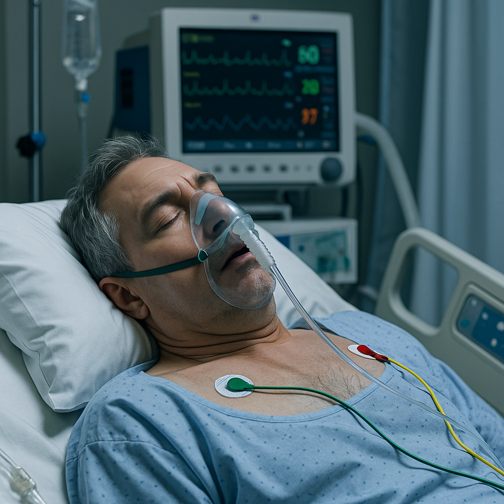
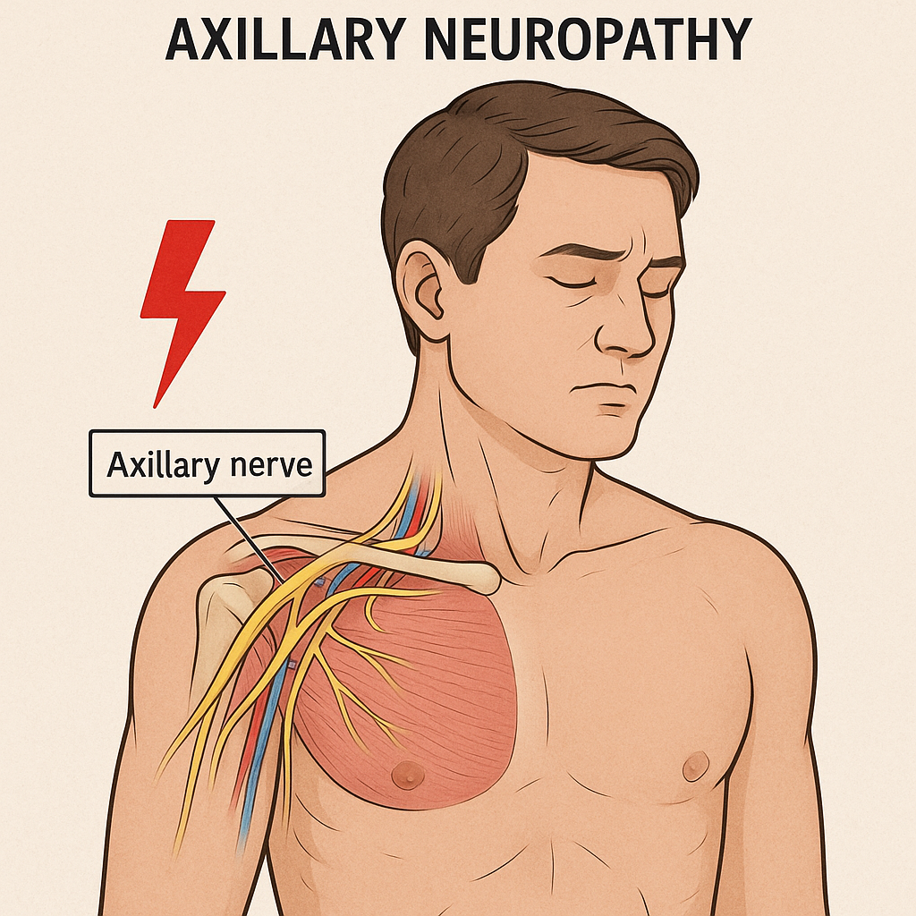
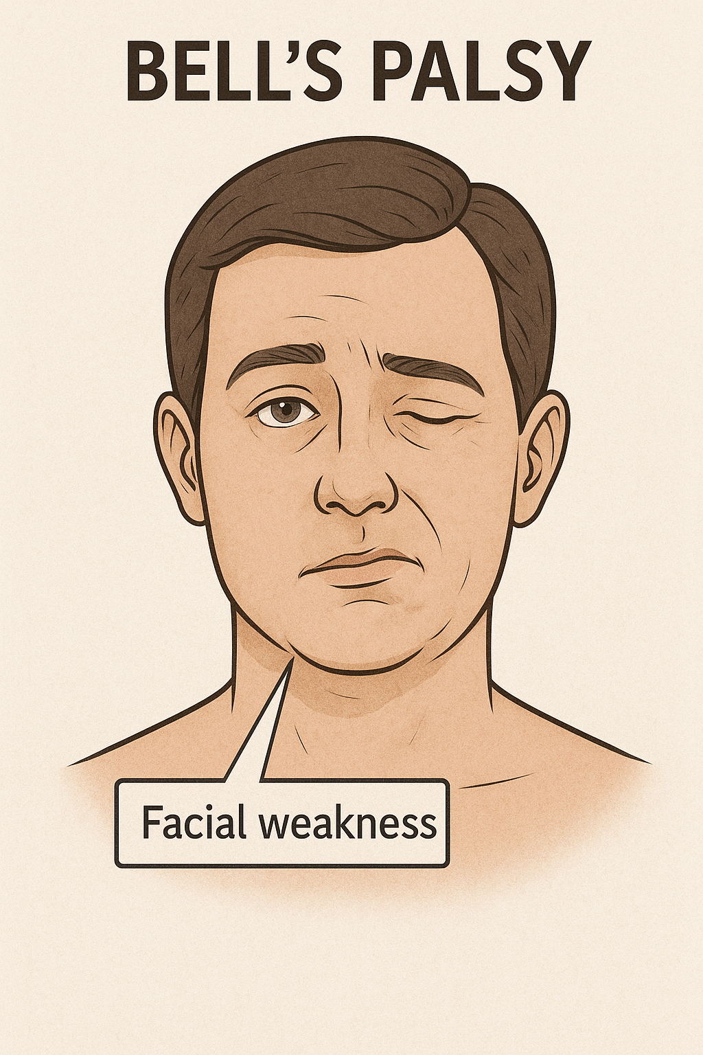
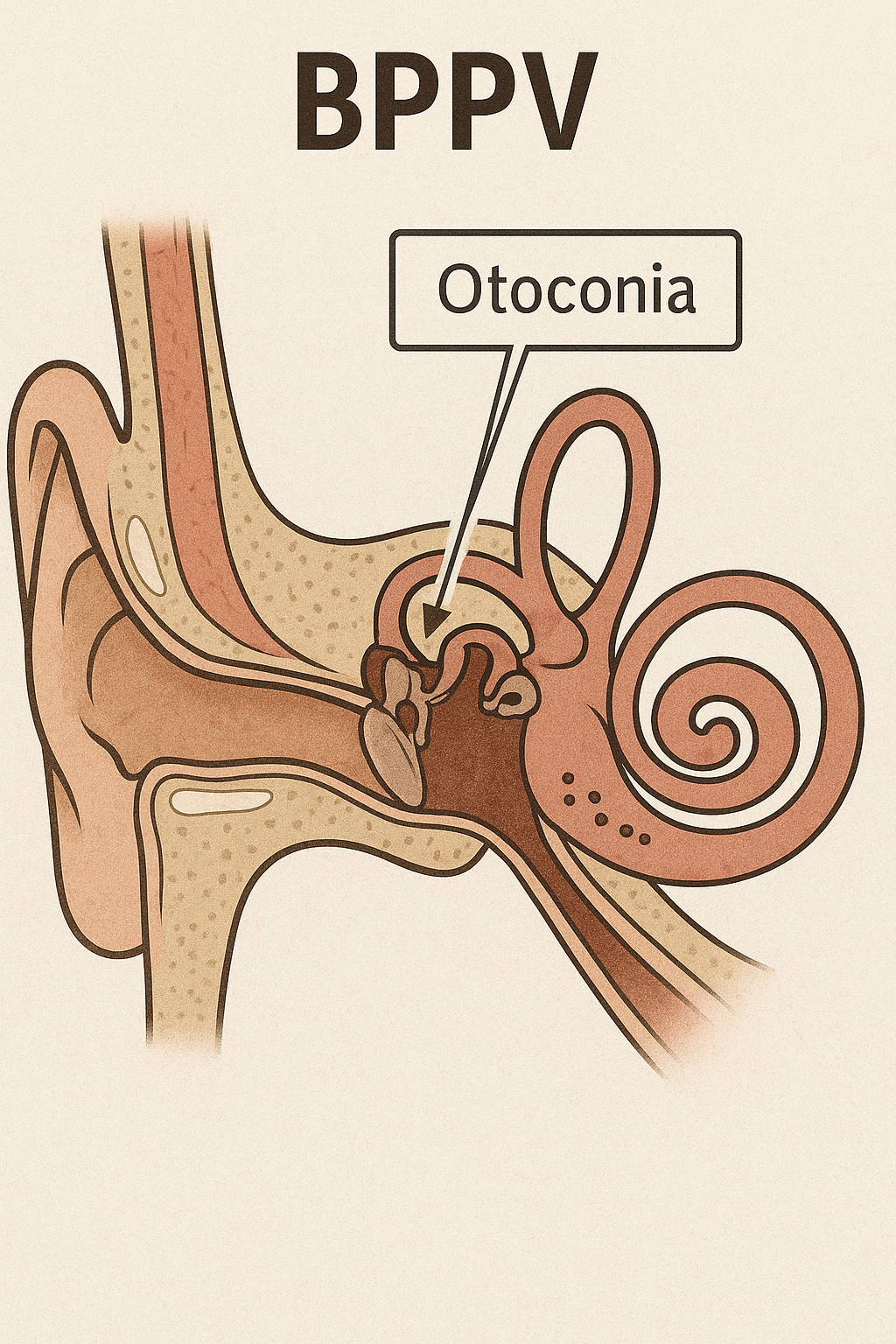
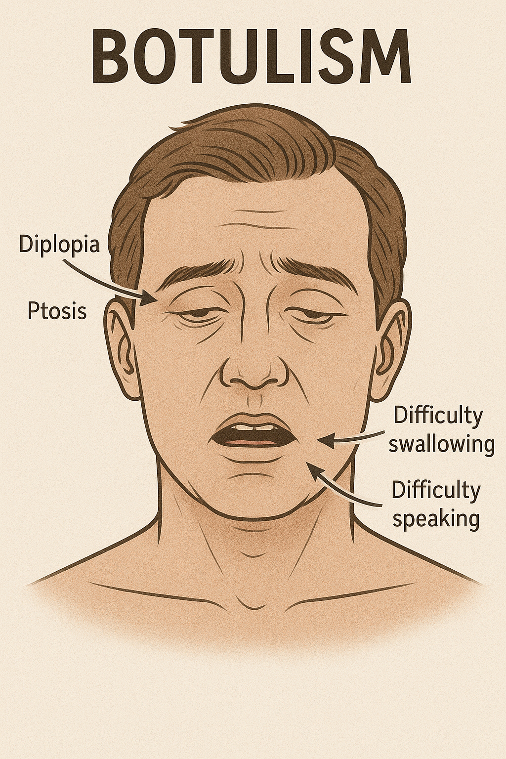
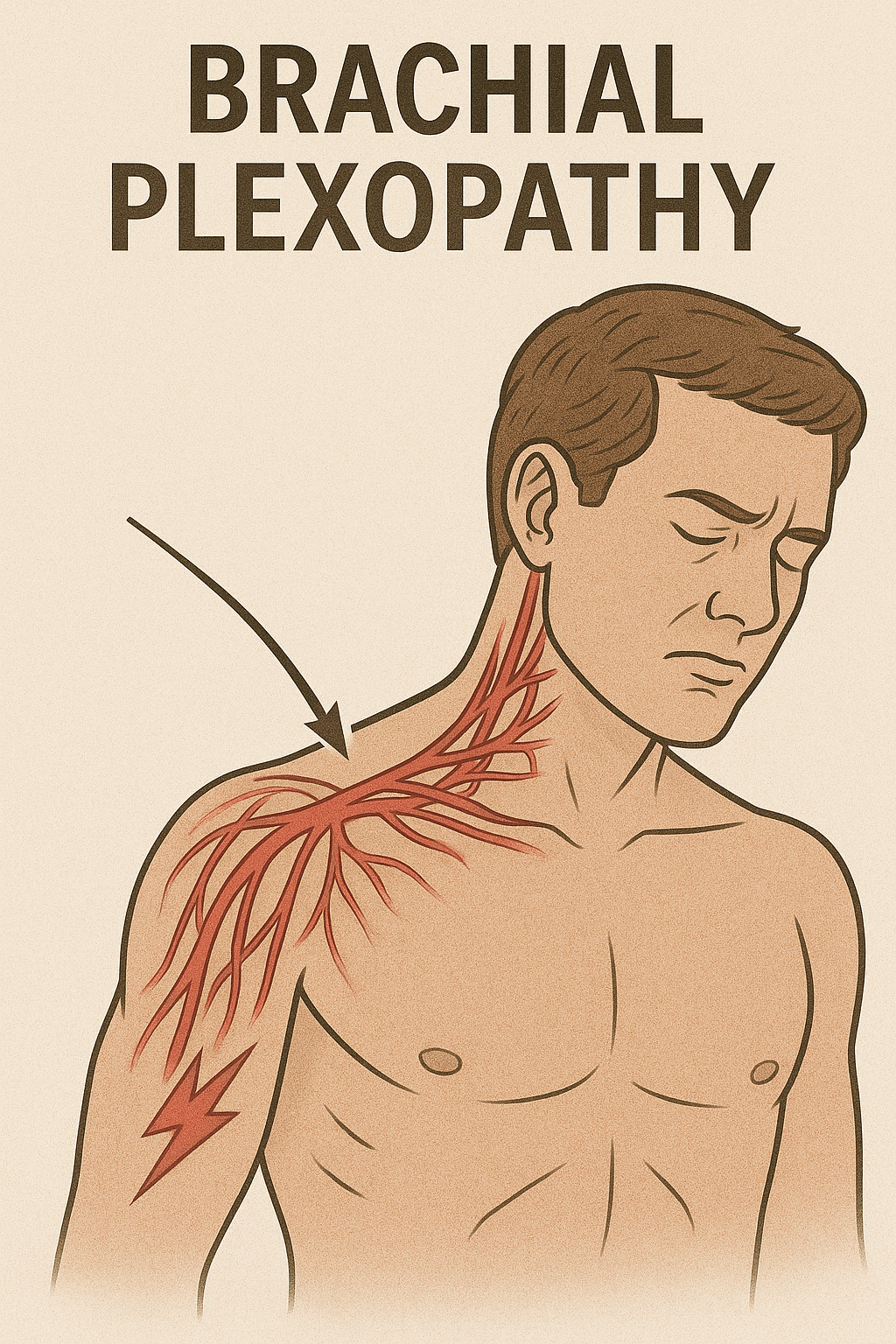
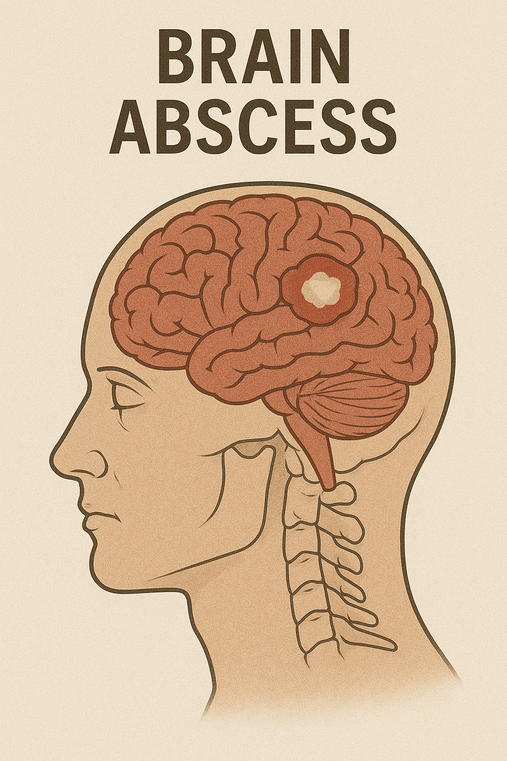
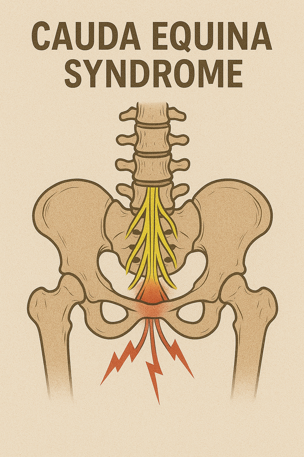
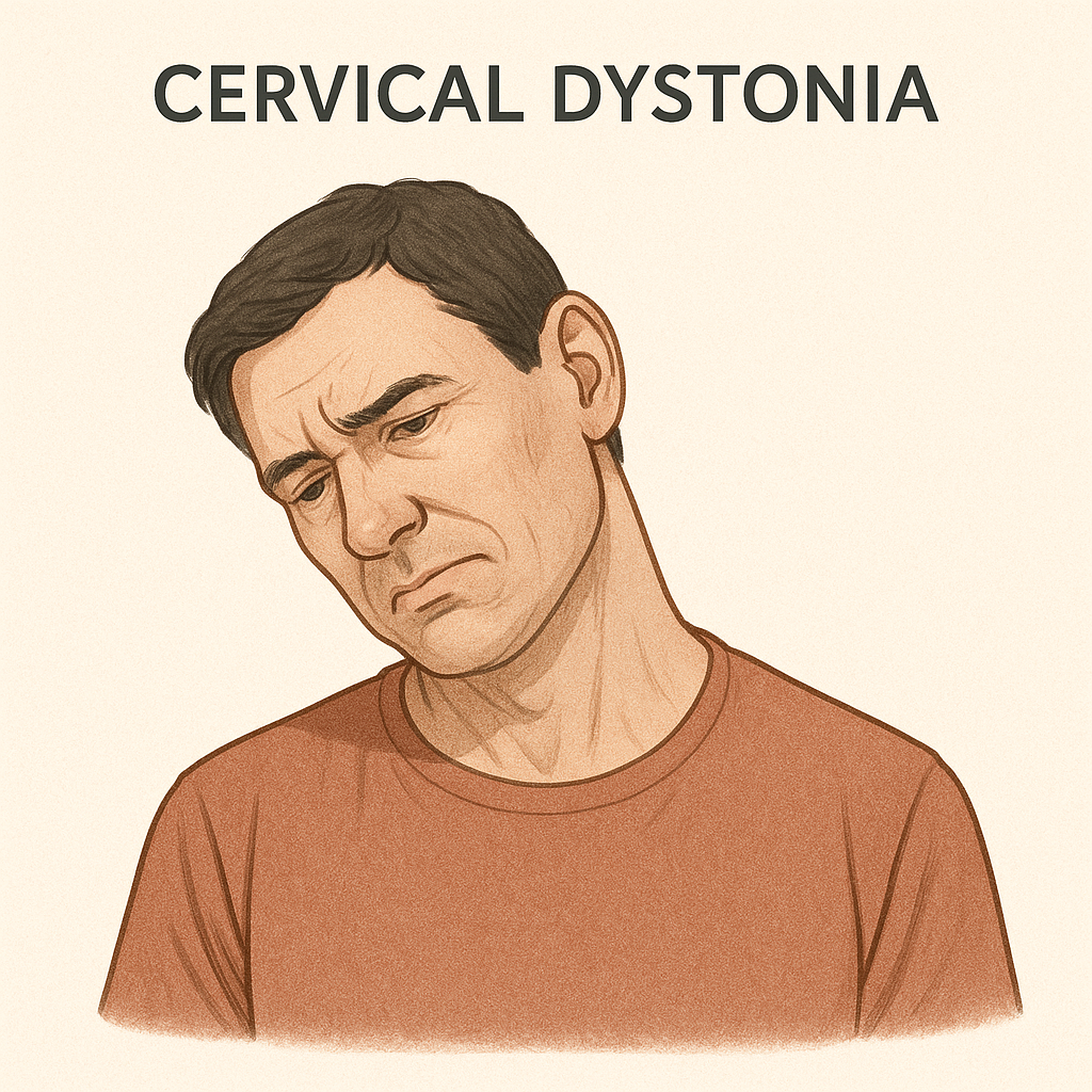
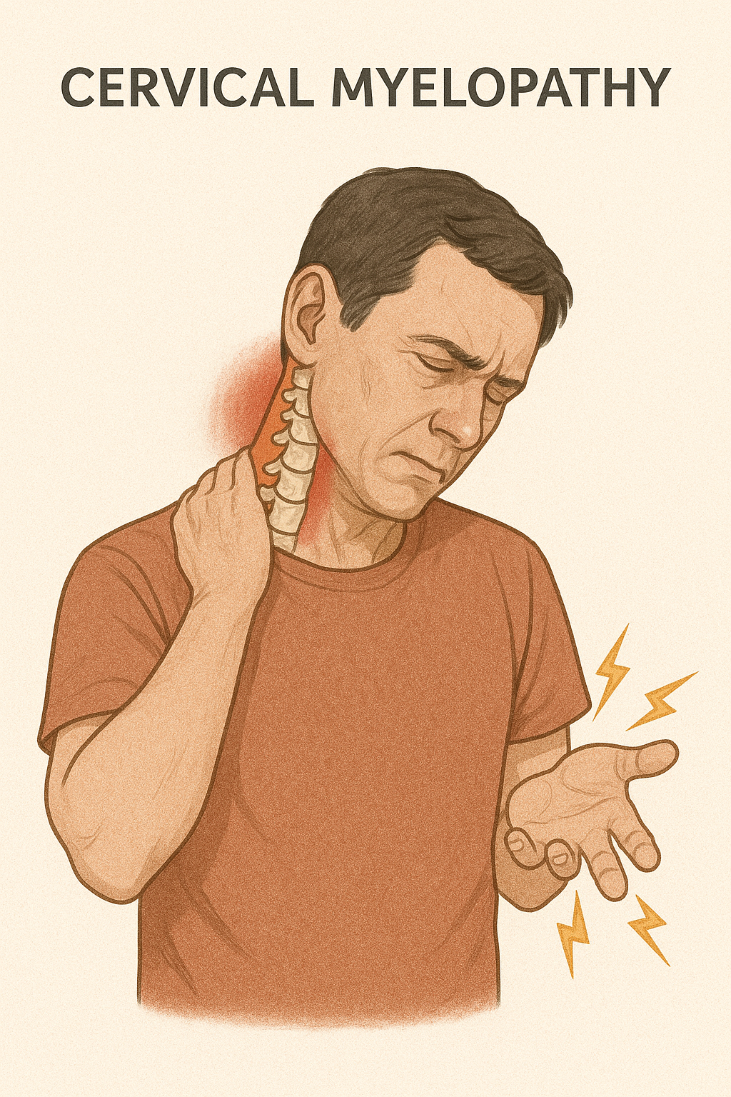
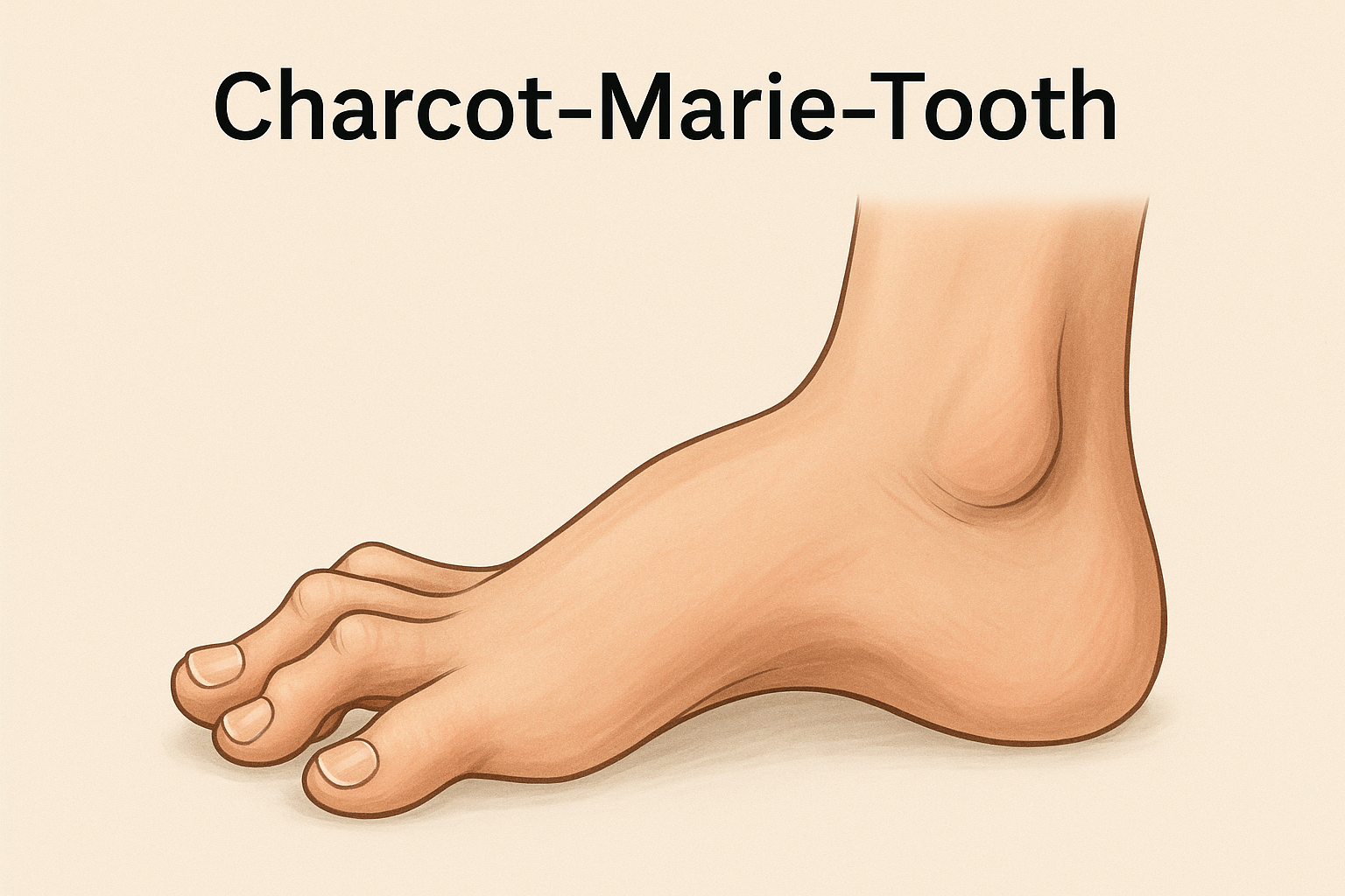
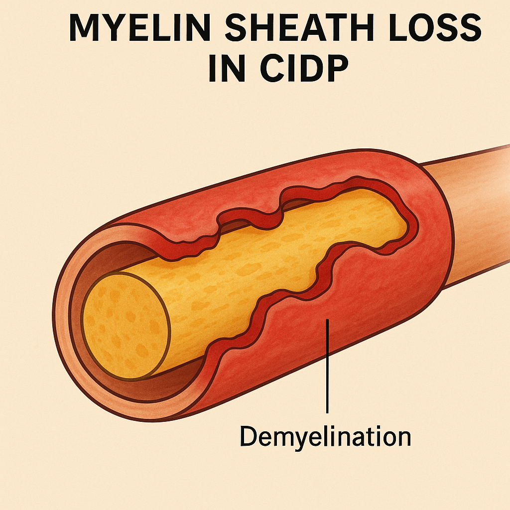
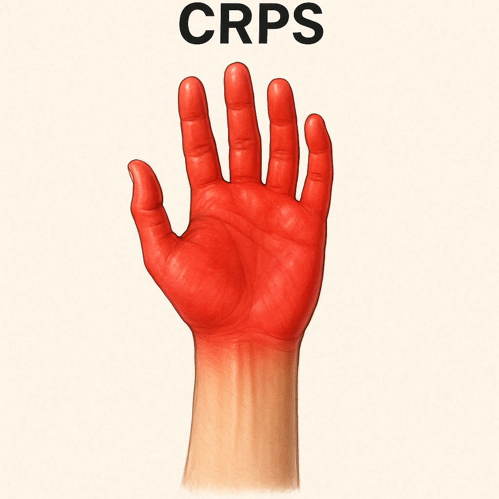
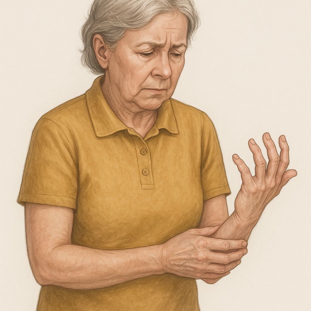
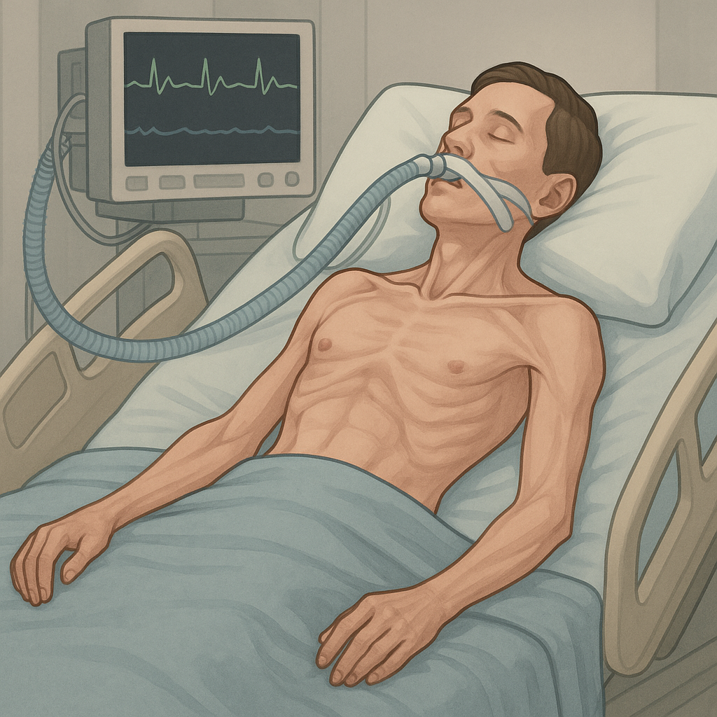
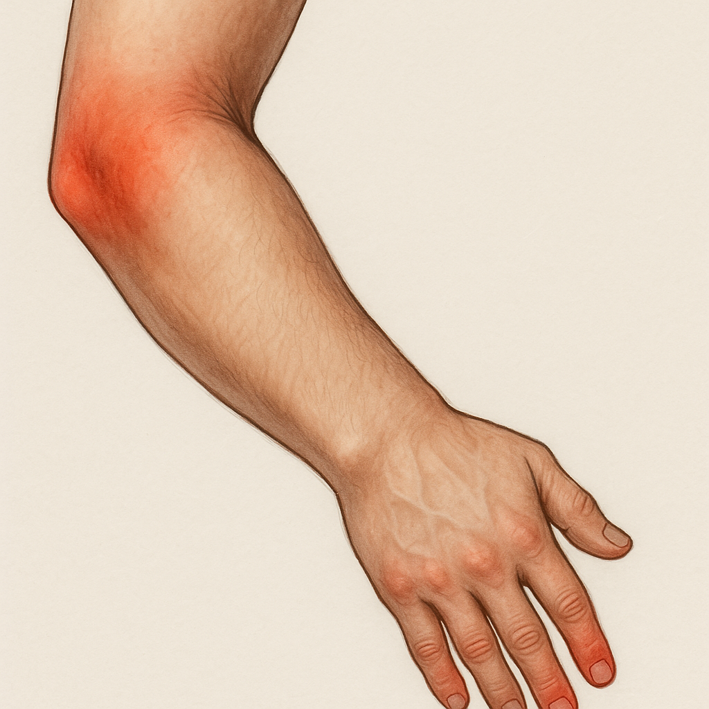
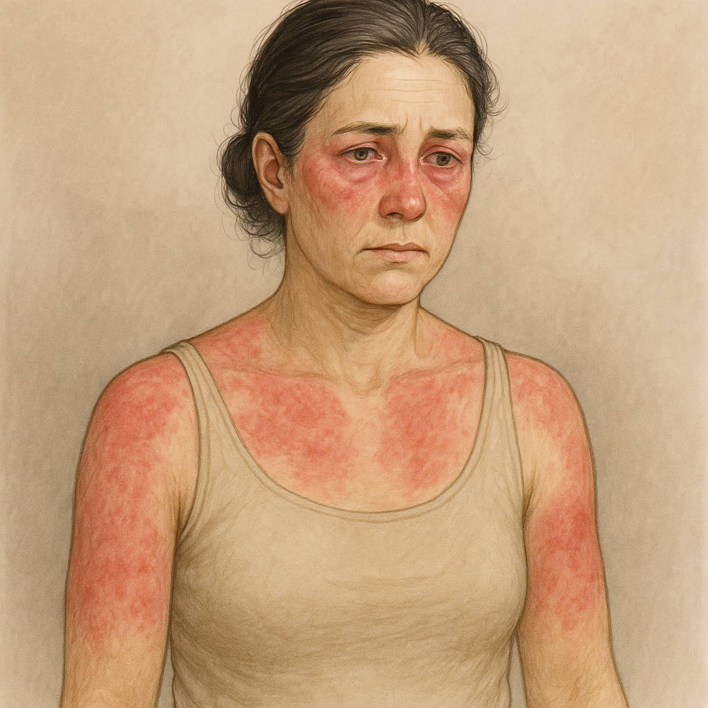
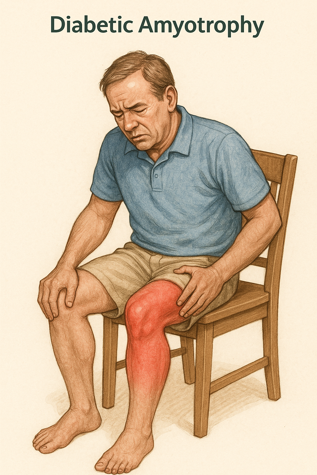
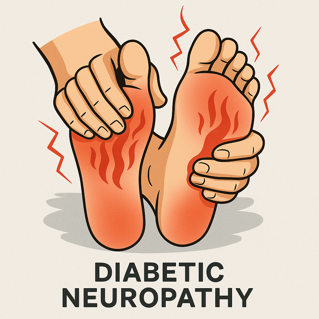
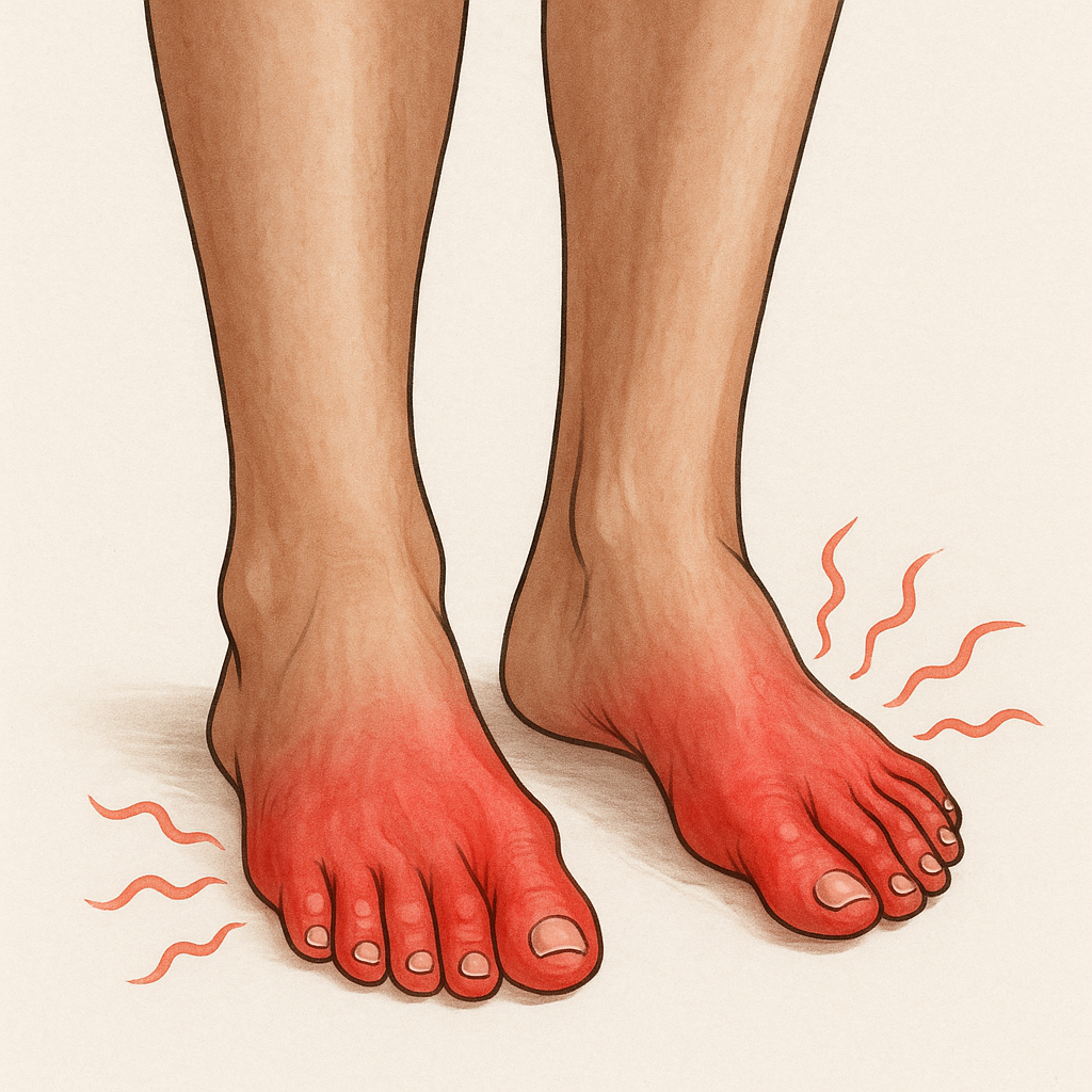
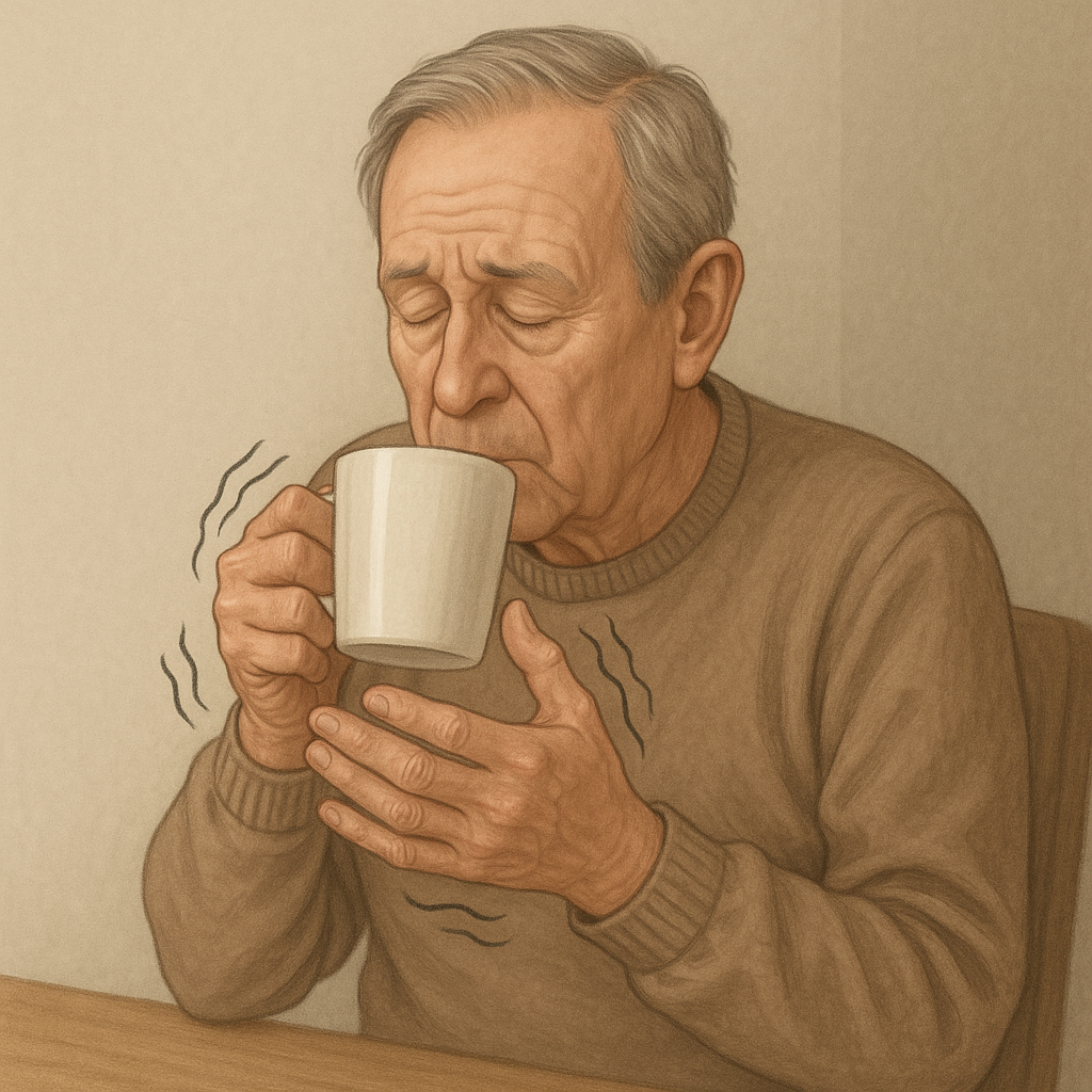
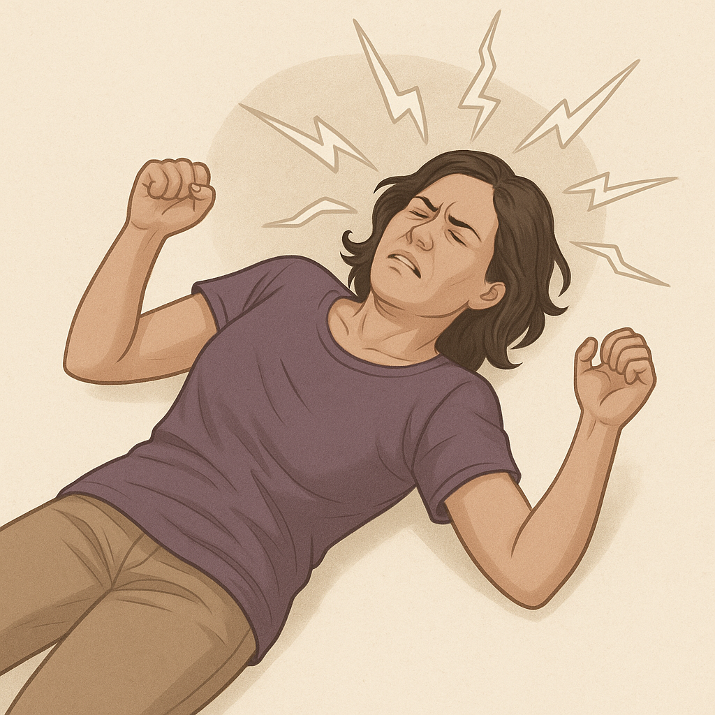
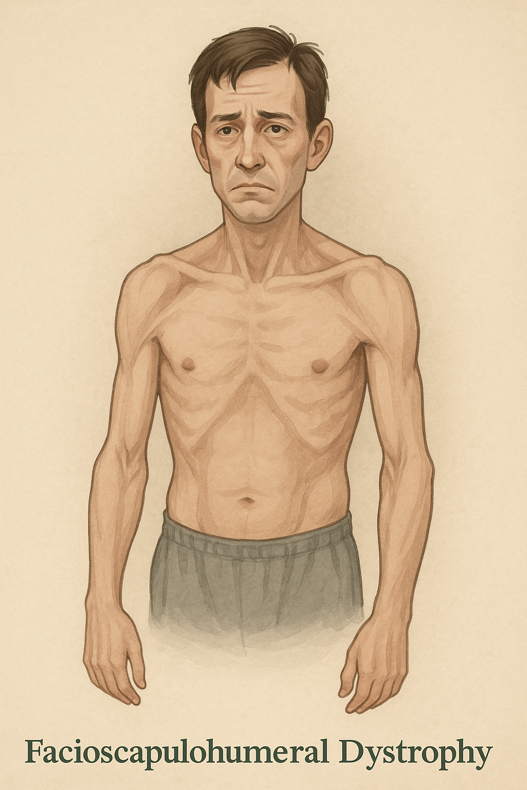
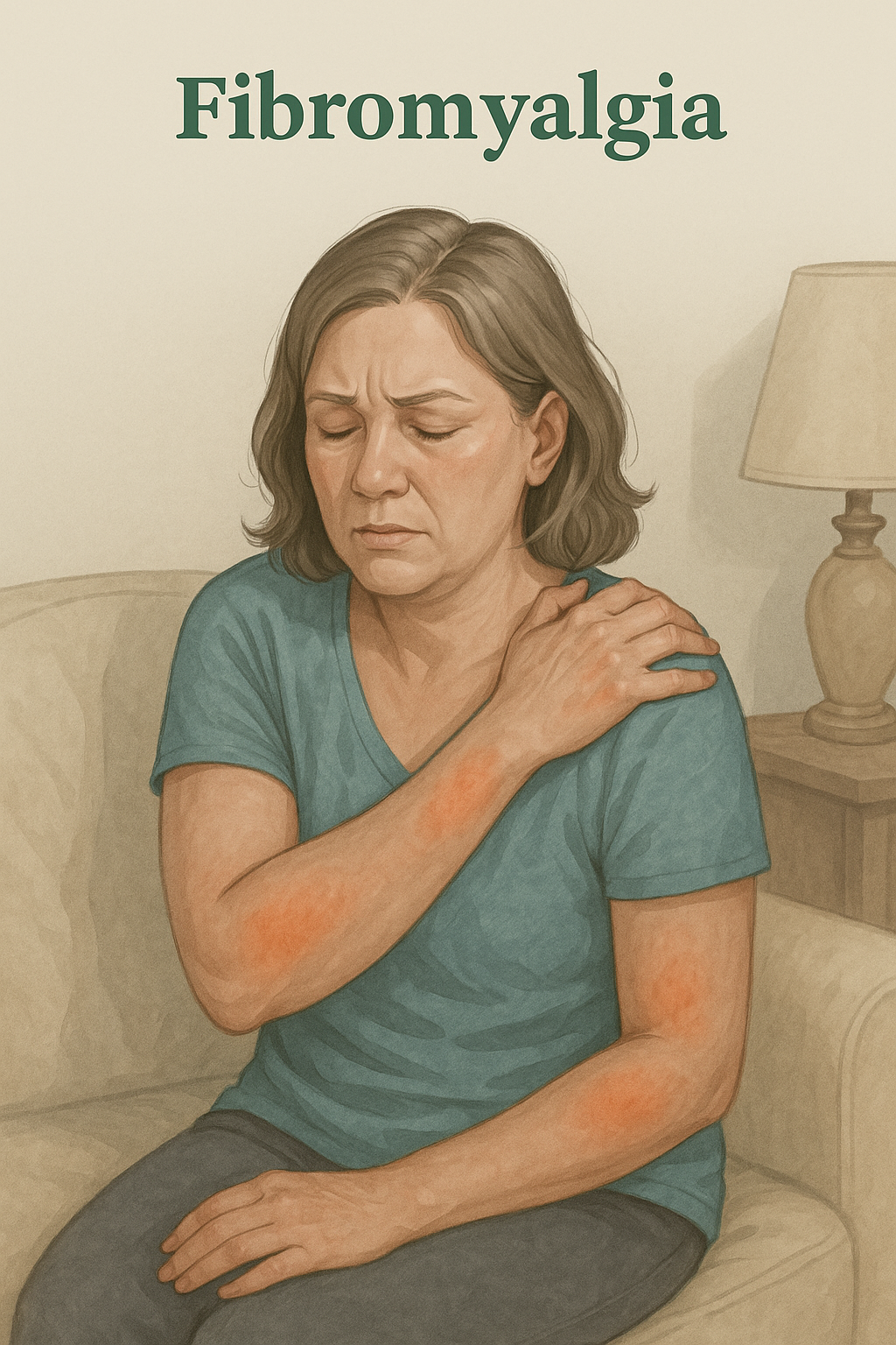
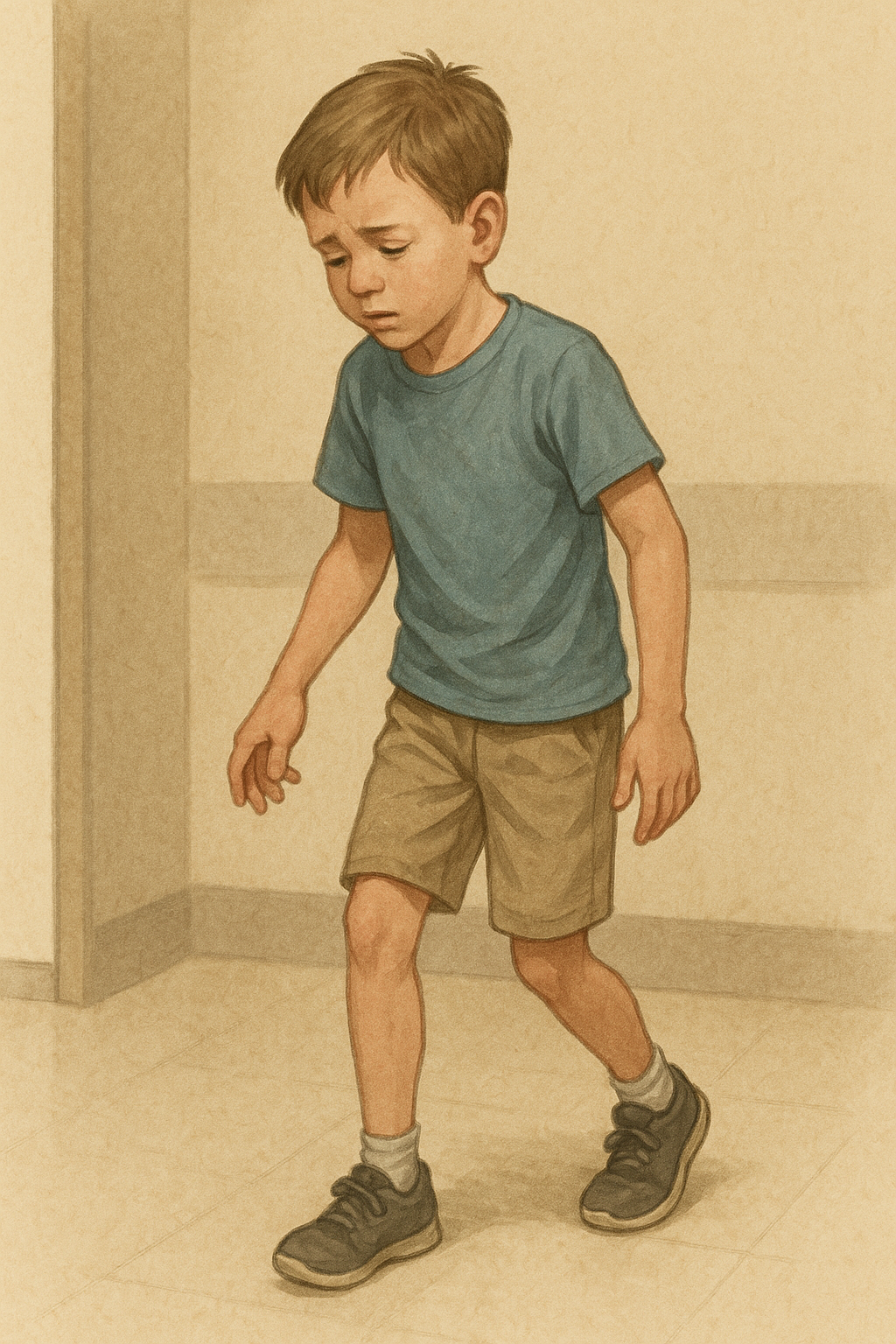
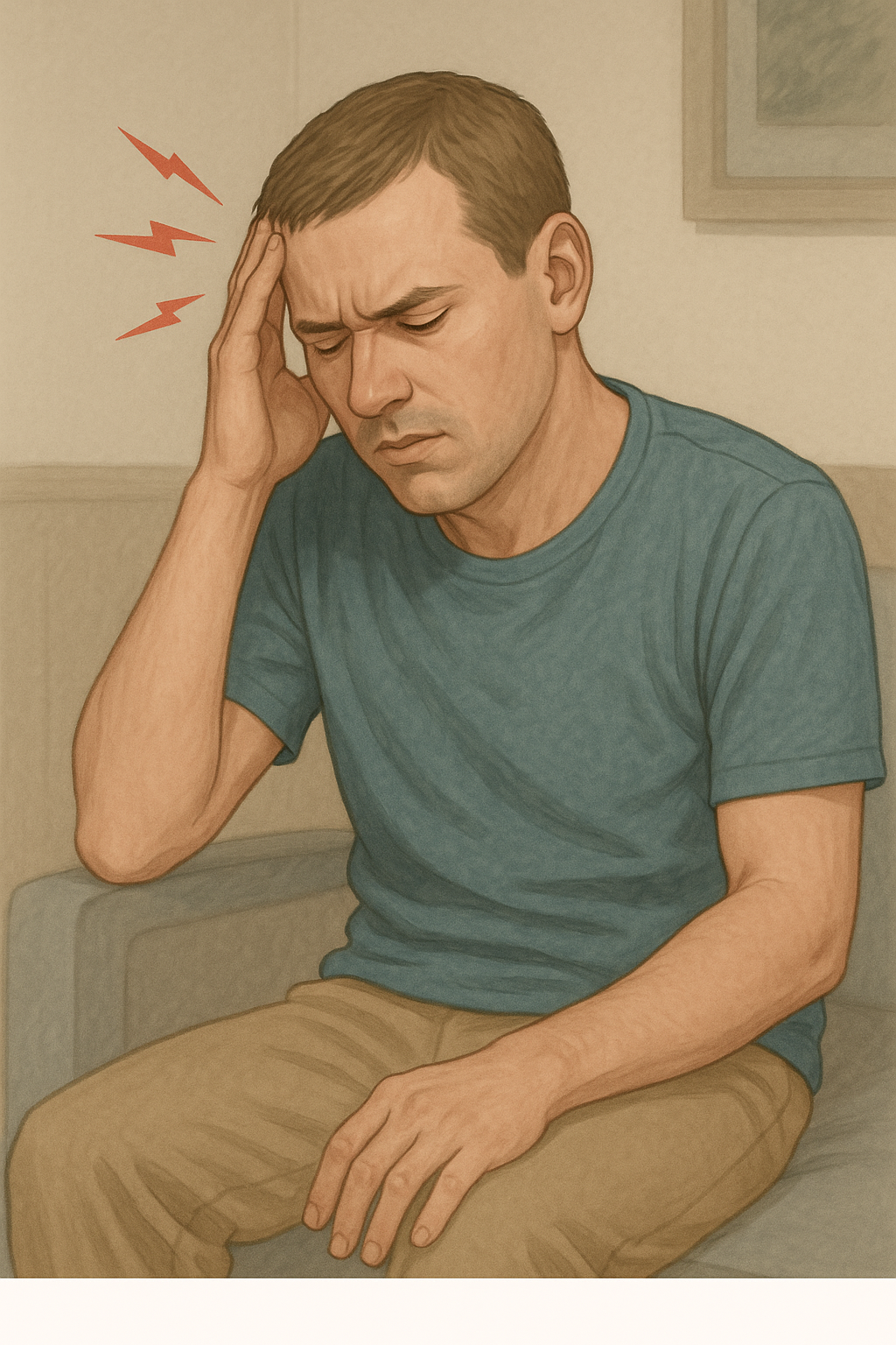
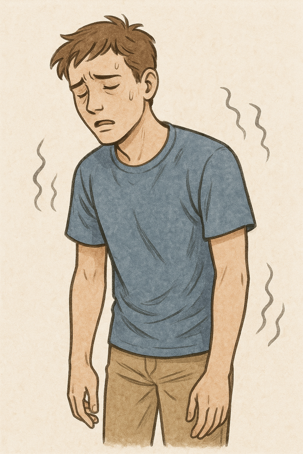
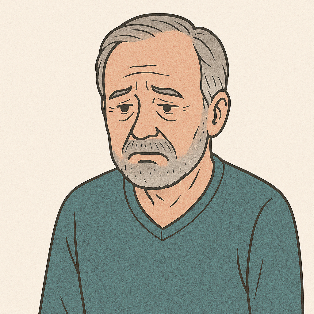
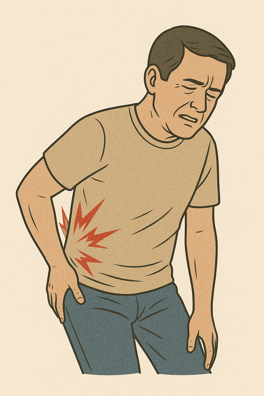
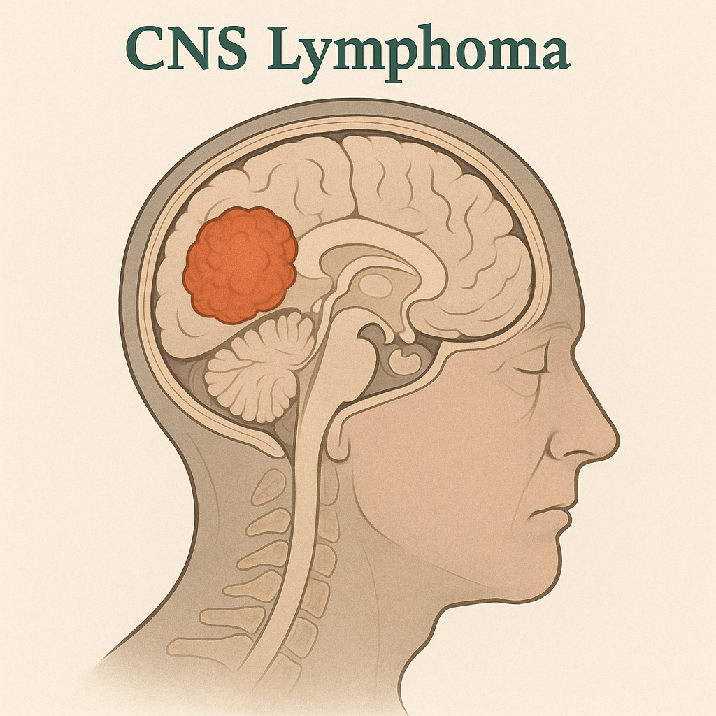
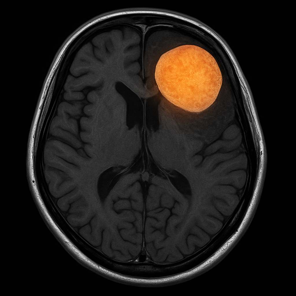
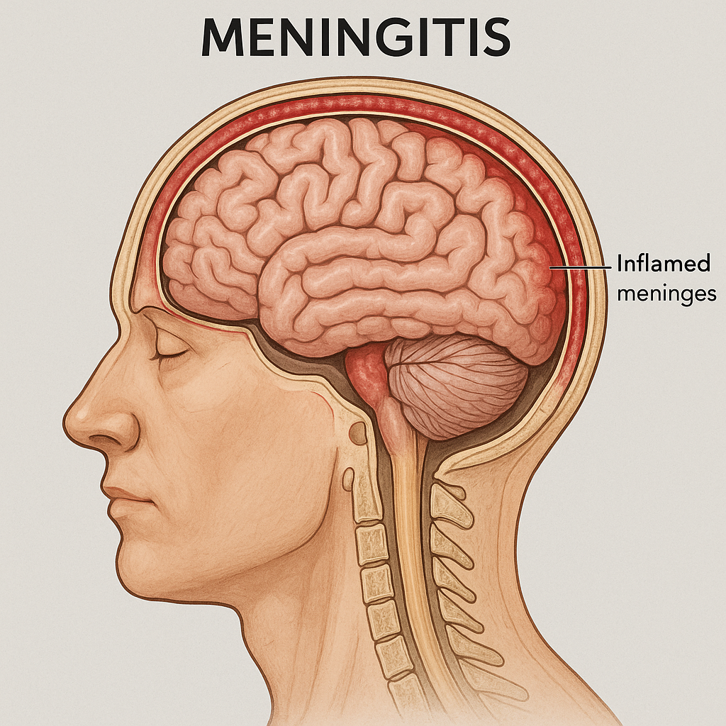
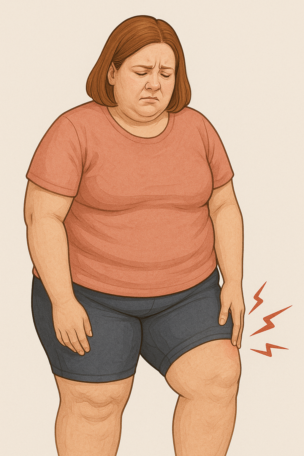
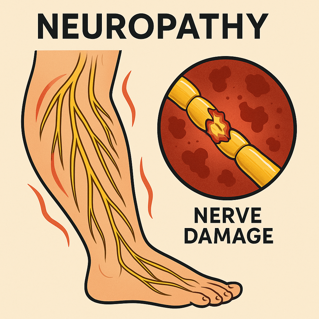
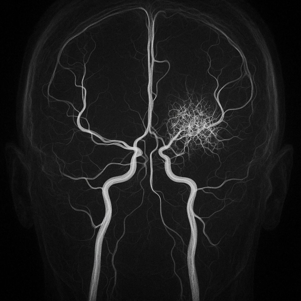
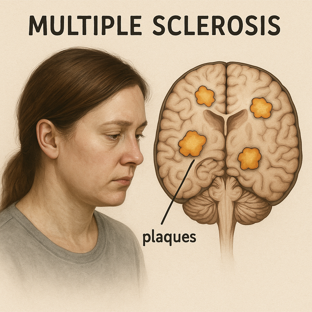
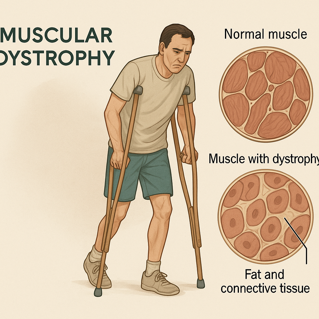
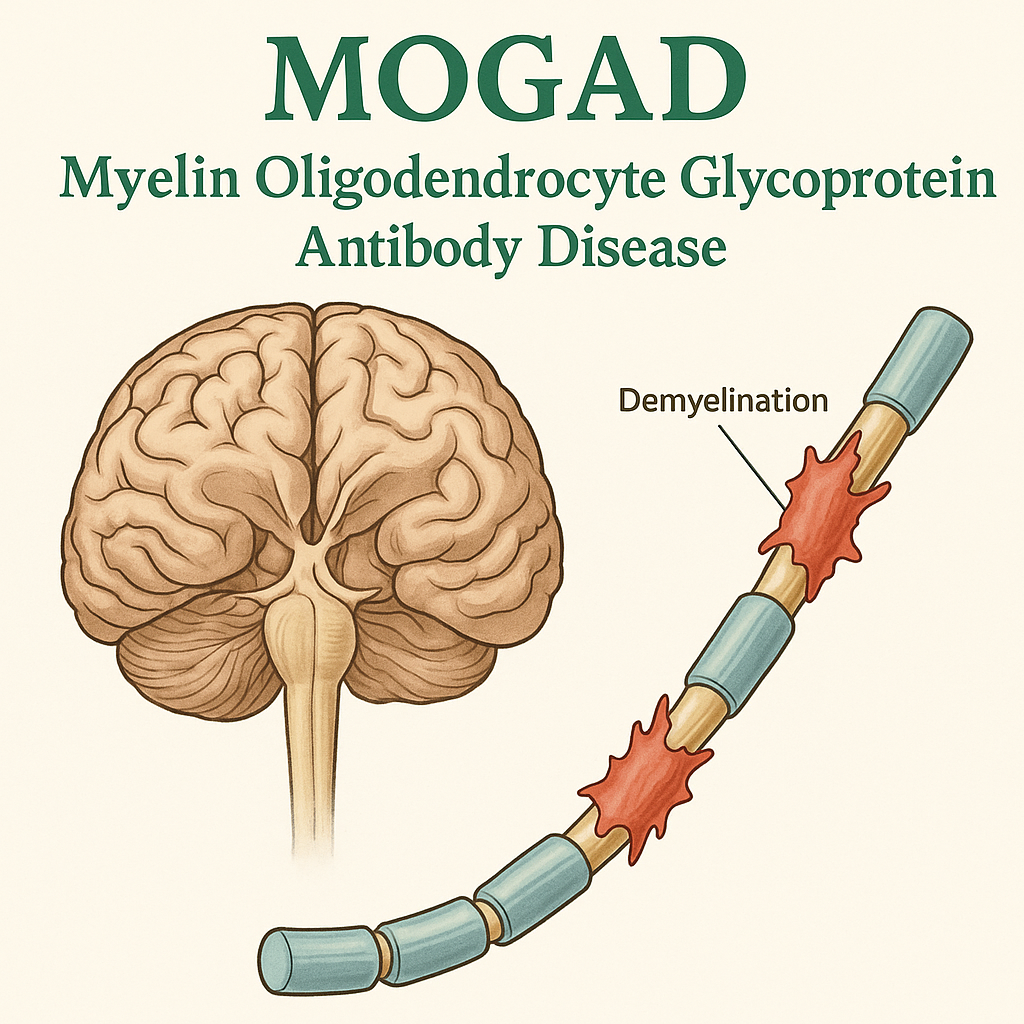
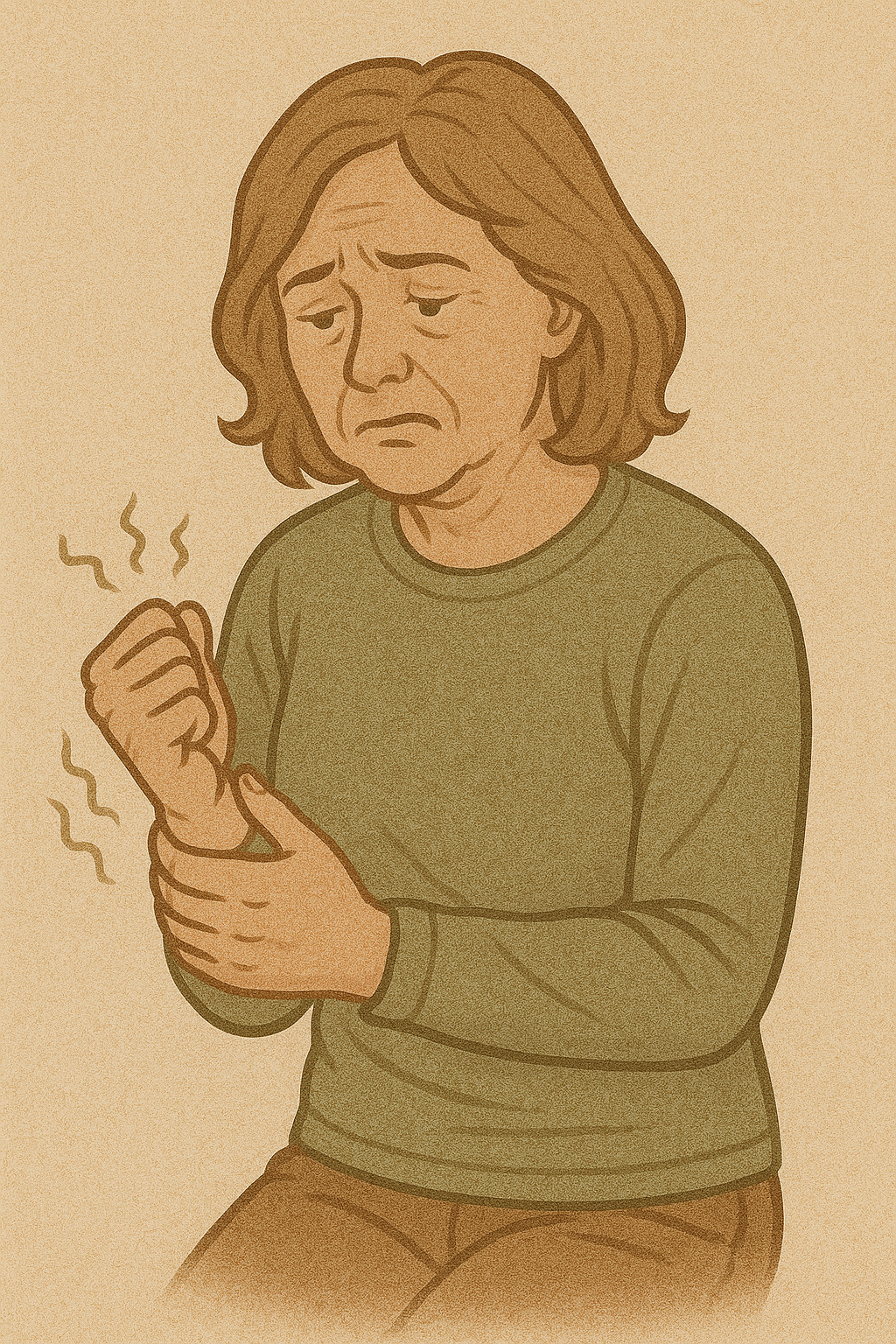
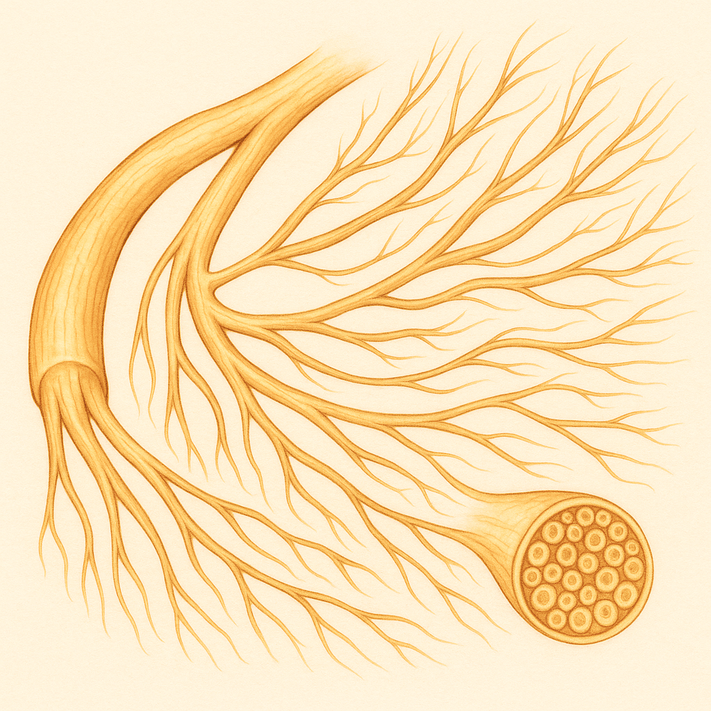
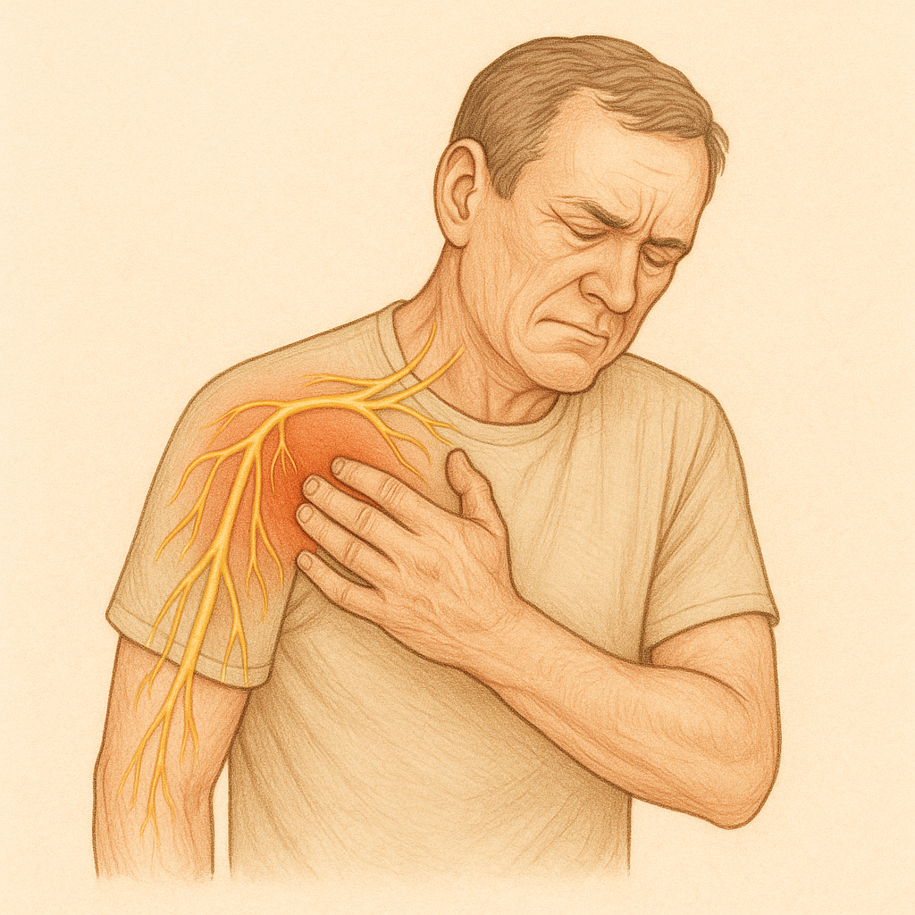
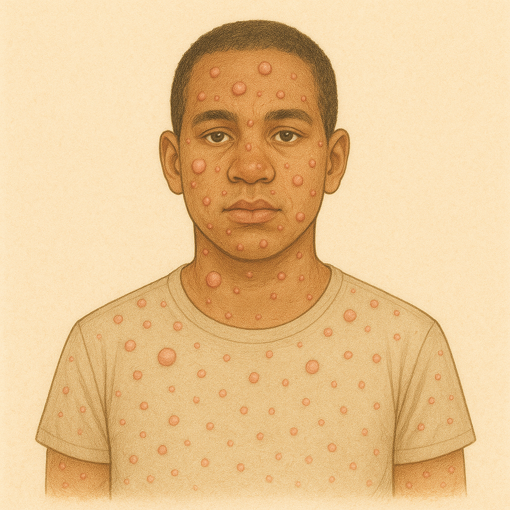
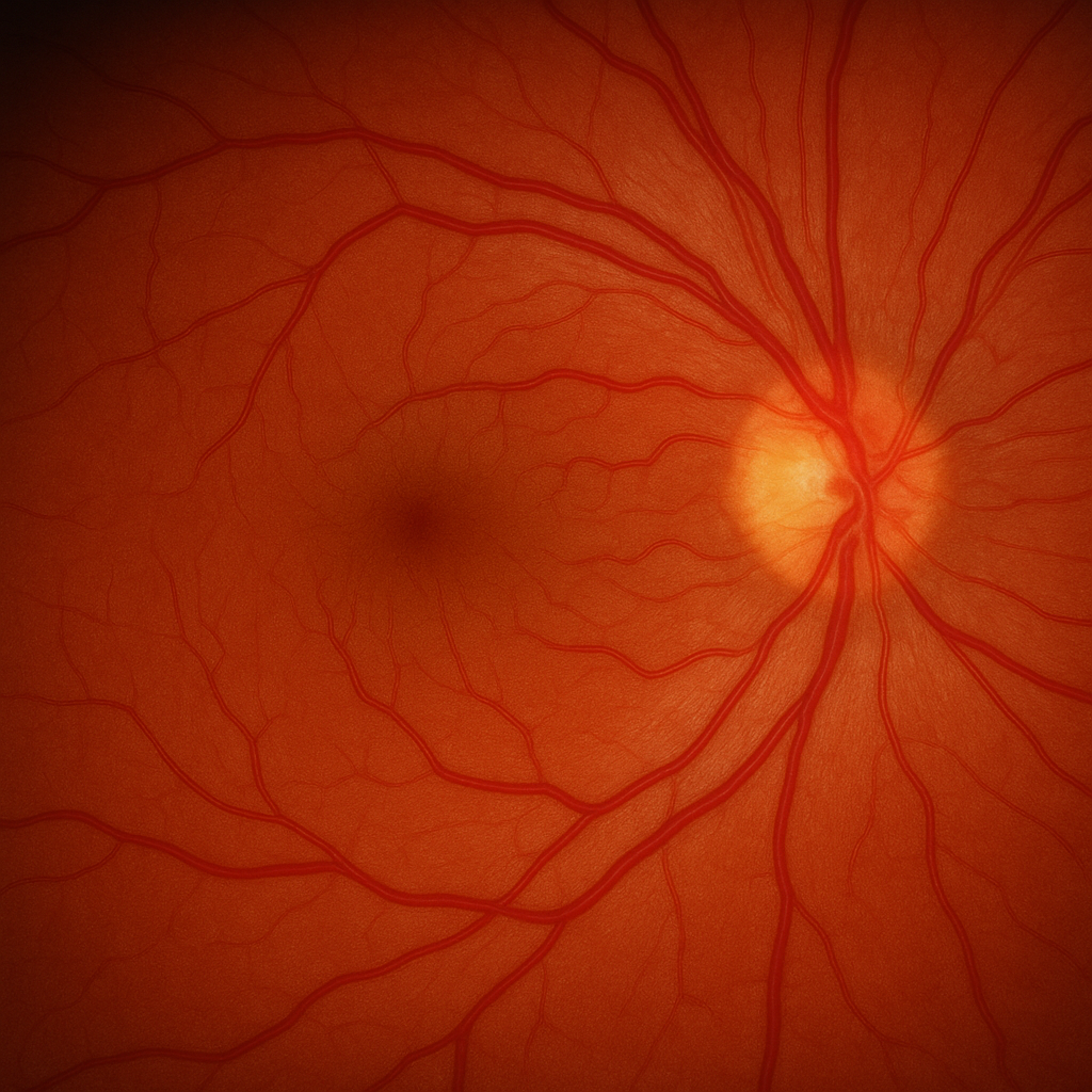
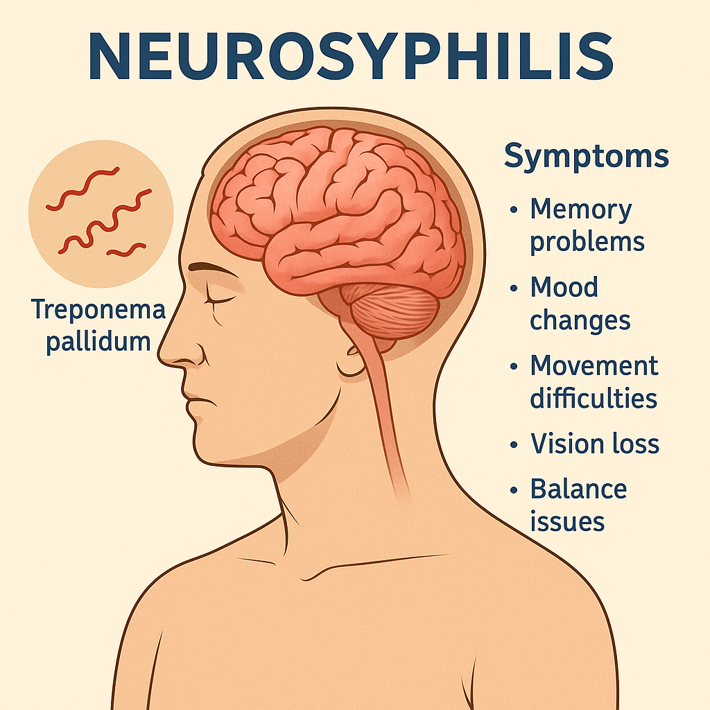
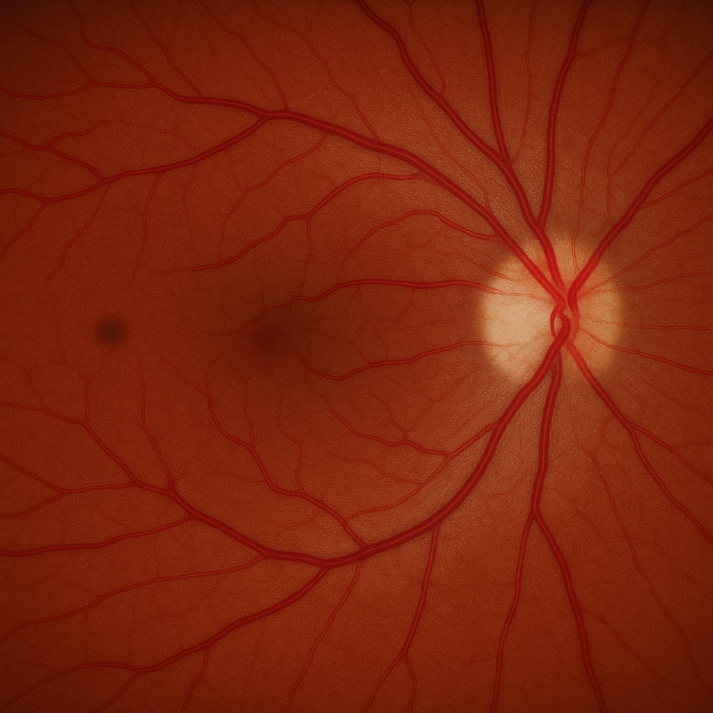
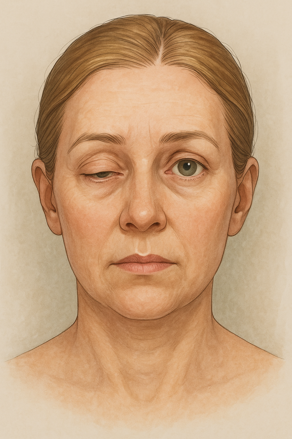
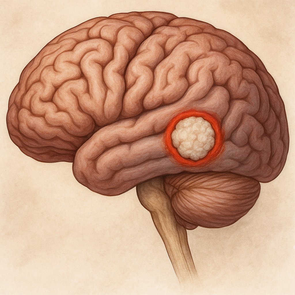
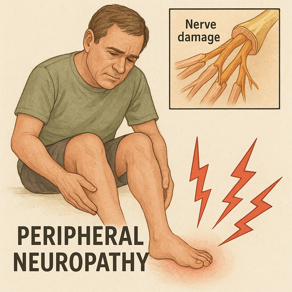
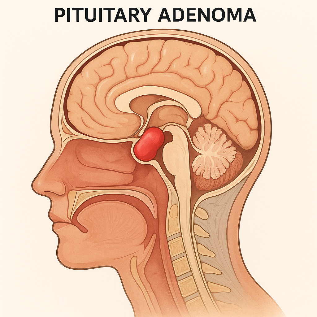
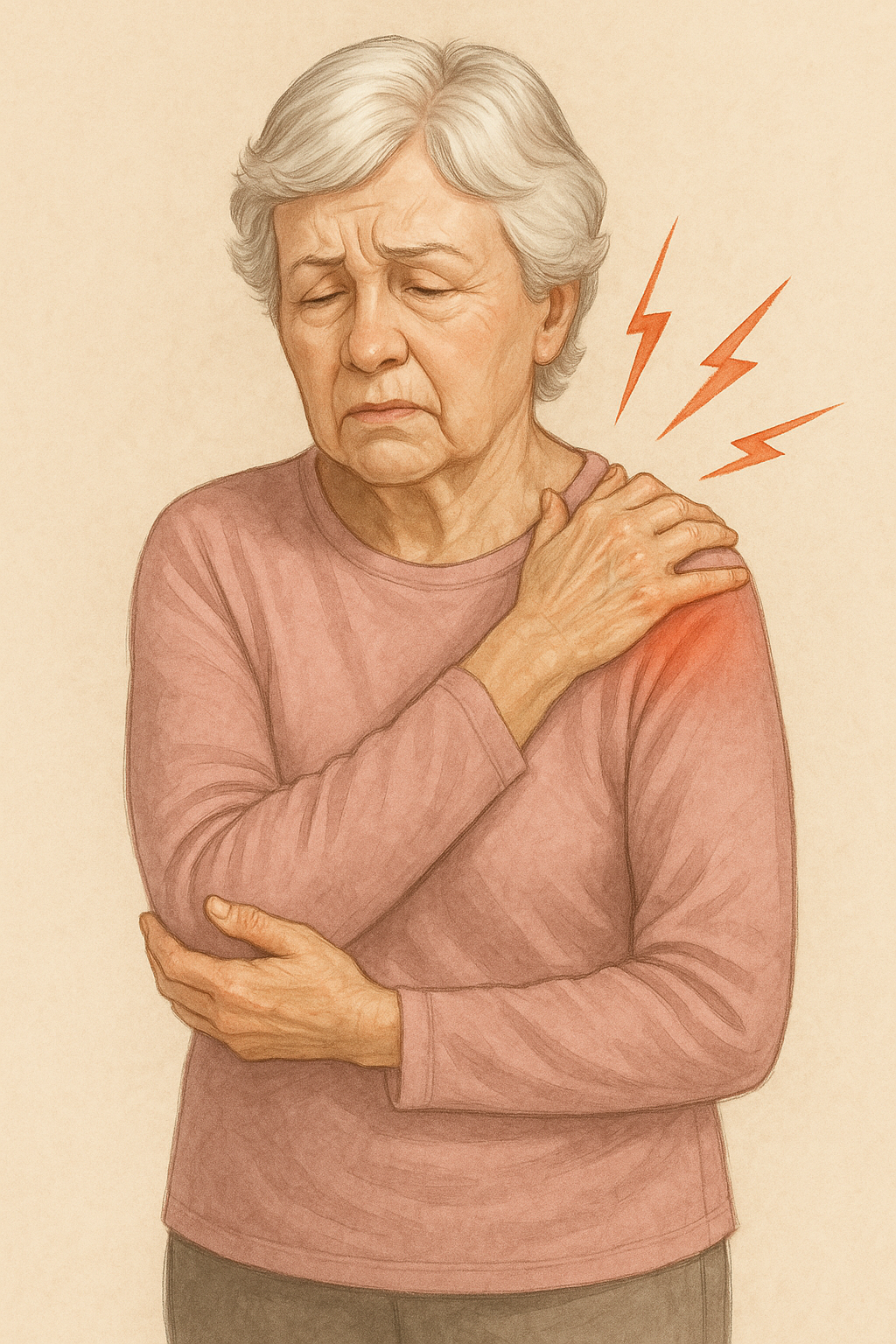
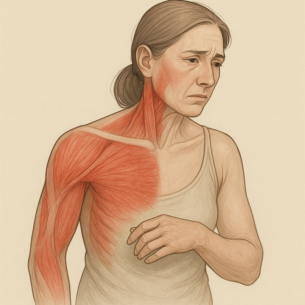
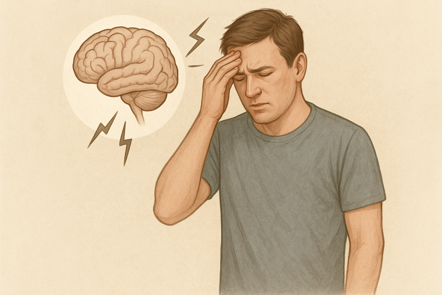
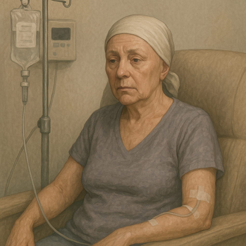
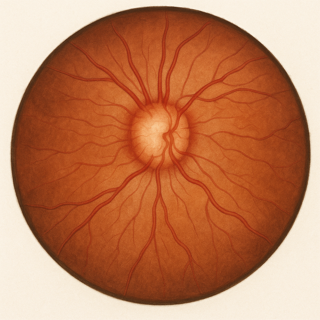
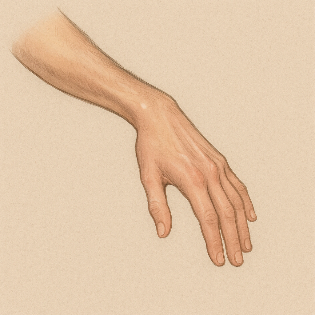
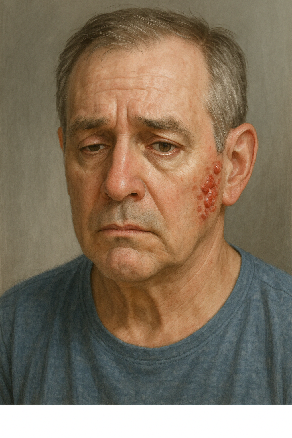
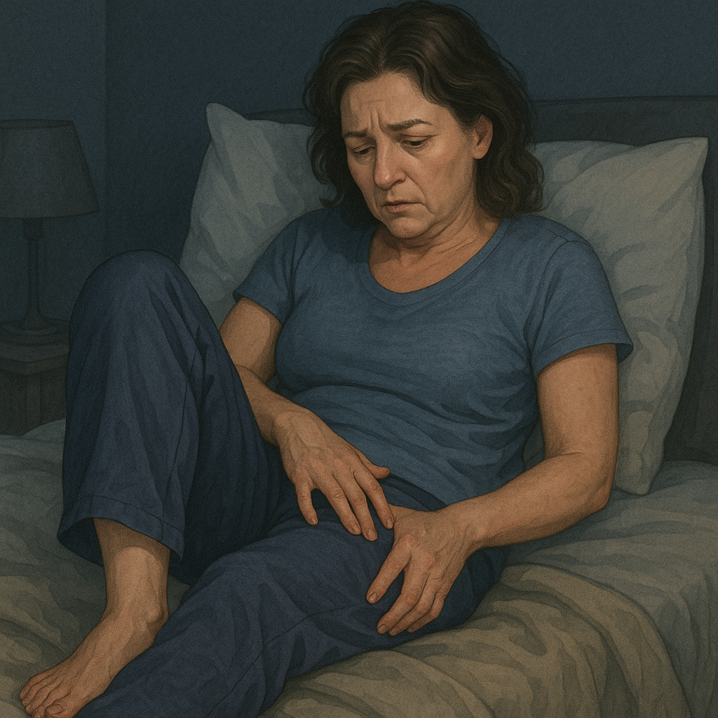
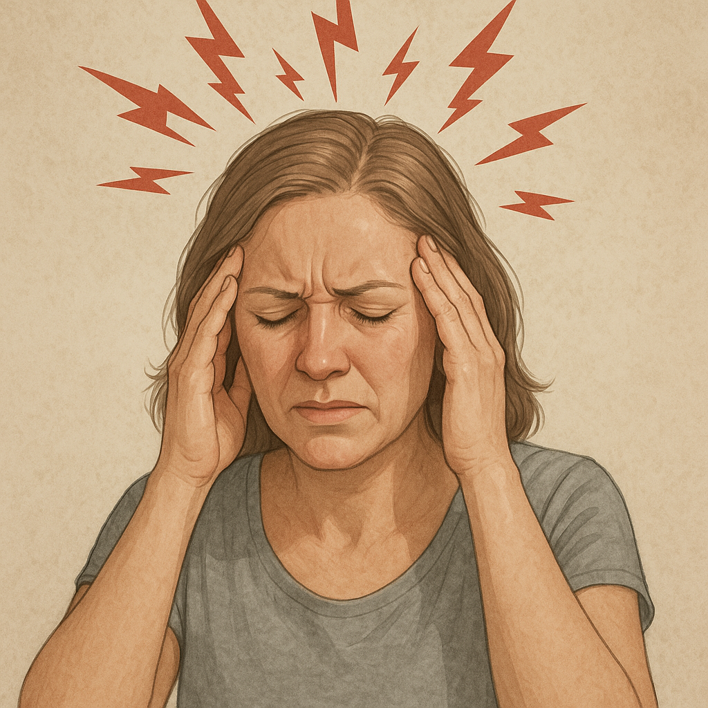
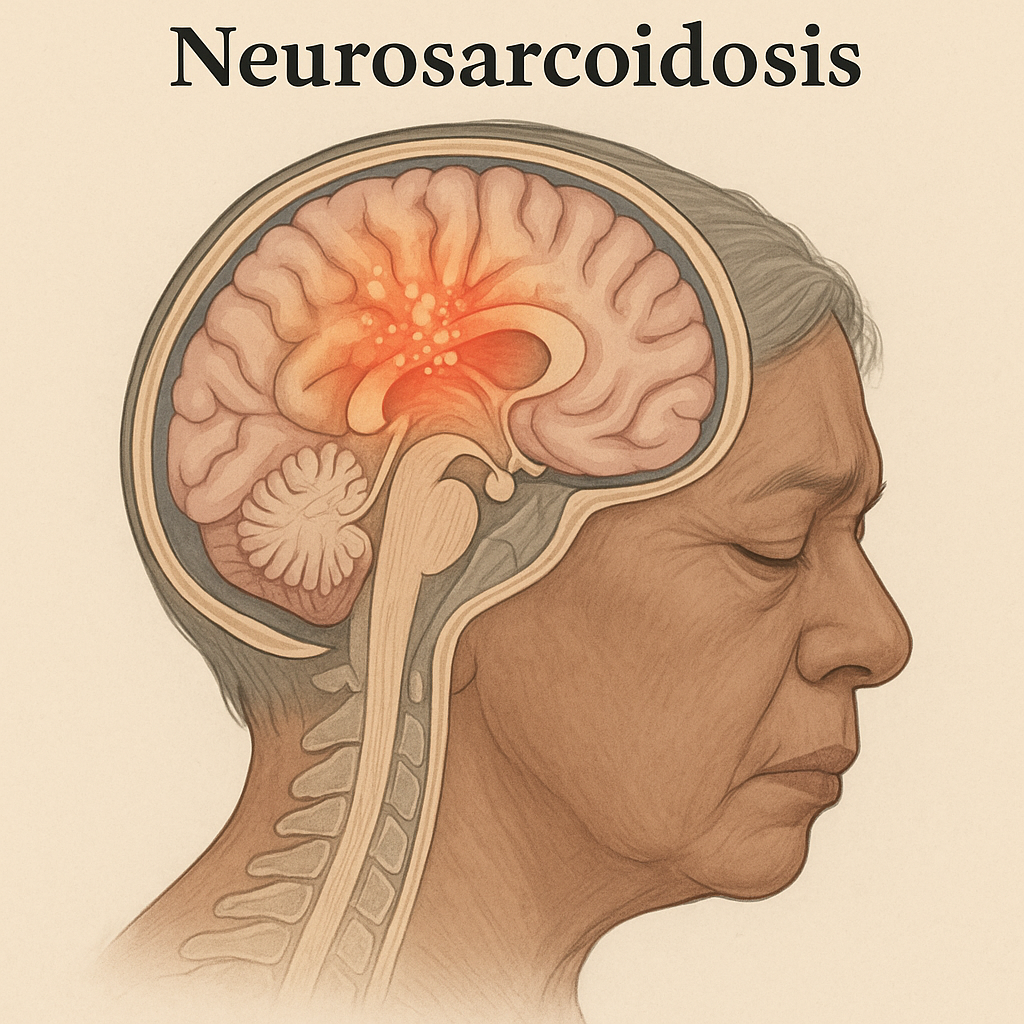